Unlocking the Mystery of Tachycardia
Mechanisms Behind the Beat
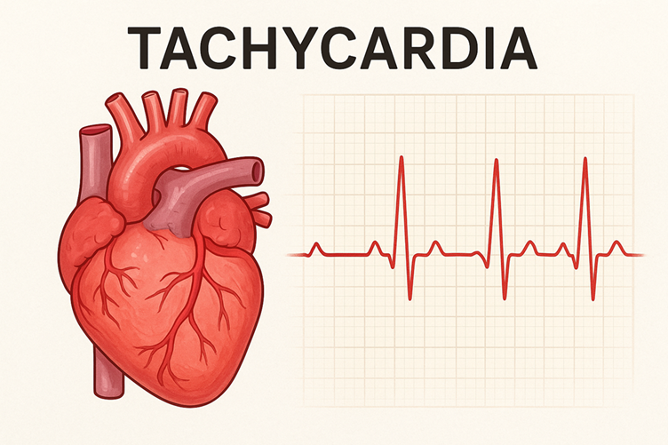
A medical disorder known as tachycardia is typified by an unusually elevated heart rate. A resting heart rate exceeding 100 beats per minute (bpm) is considered its definition.
While prolonged occurrences of other types of tachycardia can result in heart failure, some types of tachycardia, especially ventricular tachycardia (VT), are potentially fatal and can cause abrupt cardiac arrest. Sinus tachycardia, which is frequently a typical reaction to stress or activity, may not be harmful, though. Any new or unexpected rapid heart rate requires immediate medical attention, particularly if it is accompanied by dizziness, fainting, chest pain, or shortness of breath.
You’ll encounter two basic electrical problems: increased automaticity and re-entry circuits. Automaticity means a focus—sinus node, atrial ectopic focus, or an irritable ventricular focus—fires faster than normal; sinus tachycardia typically sits just above 100 bpm, while atrial tachycardia often produces rates of 120–180 bpm. Re-entry happens when electrical signals loop back on themselves; examples include AV nodal re-entrant tachycardia (AVNRT) and various types of paroxysmal supraventricular tachycardia
Ventricular tachycardia (VT) arises from the ventricles and can be monomorphic (stable QRS morphology) or polymorphic (changing QRS, including torsades de pointes). Sustained VT is defined as lasting >30 seconds or causing hemodynamic instability. Pulseless VT requires immediate cardiopulmonary resuscitation (CPR) and defibrillation per ACLS protocols.
How Clinicians Differentiate Types of Tachycardia
Based on the location of the aberrant electrical signal’s origin in the heart, tachycardia can be divided into three primary kinds. There are numerous subcategories within these groups.
1. SVT, or supraventricular tachycardia
SVT is a fast heartbeat that starts in the atria, the heart’s upper chambers, or in the atrioventricular (AV) node, which is the region that connects the ventricles and atria. A reentrant circuit, a type of “short circuit” in the electrical system of the heart, is the source of many SVTs.
SVT types include:
In adults, atrioventricular nodal reentrant tachycardia (AVNRT) is the most prevalent form of SVT. The reason for AVNRT is an irregular electrical circuit located in or near the AV node.
An additional, aberrant electrical circuit connecting the atria and ventricles is the source of atrioventricular reciprocating tachycardia (AVRT). One type of AVRT is Wolff-Parkinson-White (WPW) syndrome.
A fast rhythm that originates from an irregular electrical location in the atria, apart from the heart’s natural pacemaker, is known as atrial tachycardia.
The most prevalent severe arrhythmia, atrial fibrillation (AFib), is typified by erratic and disorganized electrical signals in the atria.
Similar to AFib, atrial flutter is characterized by more ordered electrical signals in the atria that produce a regular but extremely rapid beat.
2. Ventricular Tachycardia (VT)
A potentially fatal rapid heartbeat that begins in the ventricles, the heart’s lower chambers, is known as ventricular tachycardia. It may hinder the ventricles’ ability to fill correctly, which would result in the body receiving insufficient blood flow.
VT types are frequently categorized based on how they show up on an electrocardiogram (ECG) and how long they last:
Monomorphic VT: Every heartbeat appears identical on an ECG, suggesting that the ventricles are the source of all electrical activity.
Polymorphic VT: There is variation in the electrical waves, indicating that the rhythm originates from different parts of the ventricles.
Pointes Torsades: a particular type of polymorphic VT that is associated with a prolonged QT interval and can be fatal.
Sustained VT: Hemodynamic instability or an arrhythmia lasting more than 30 seconds.
Non-sustained VT (NSVT): The arrhythmia resolves on its own after less than 30 seconds.
3. Sinus Tachycardia
Sinus tachycardia is a typical rise in heart rate that starts in the sinus node, the heart’s natural pacemaker. It is usually the body’s normal reaction to a stimulus and is not regarded as an arrhythmia.
You’ll see ECG clues: narrow-complex tachycardia (QRS <120 ms) usually indicates supraventricular origin; wide-complex tachycardia (QRS ≥120 ms) raises suspicion for VT. Adenosine (6 mg, then 12 mg if needed) can terminate or unmask AVNRT/AVRT but must be avoided in suspected VT and atrial fibrillation with accessory pathways. The REVERT trial showed a modified Valsalva maneuver converted SVT to sinus rhythm in about 43% of cases versus 17% with the standard technique—an actionable tip you can use in stable episodes.
Orthostatic testing helps diagnose POTS: a sustained heart rate increase of ≥30 bpm within 10 minutes of standing (or ≥40 bpm in adolescents) without orthostatic hypotension. For torsades de pointes, you’ll typically see a polymorphic VT in the setting of prolonged QT; magnesium sulfate (2 g IV) is the first-line acute treatment.
Red Flags and When to Act
You must treat sudden syncope, chest pain, severe shortness of breath, hypotension, or altered mental status as emergencies—these signs suggest poor perfusion from the tachyarrhythmia. A heart rate consistently over 150 bpm with dizziness or low blood pressure often requires urgent synchronized cardioversion. Pulseless VT and ventricular fibrillation demand immediate defibrillation and ACLS measures; survival drops roughly 7–10% per minute without defibrillation.
If episodes are brief and you’re otherwise well, ambulatory ECG monitoring (24–48 hour Holter or 7–30 day event monitor) will often catch paroxysmal events during outpatient evaluation. An implantable loop recorder can be useful when symptoms are infrequent but concerning.
Conventional Treatment Pathways
You’ll typically start with vagal maneuvers for stable SVT; modified Valsalva is the most effective bedside option. Medication treatment depends on the type of rhythm: use adenosine for AVNRT/AVRT, beta-blockers or calcium-channel blockers to control the heart rate in atrial tachycardias, and antiarrhythmics like amiodar For sustained monomorphic VT after myocardial infarction, catheter ablation and implantable cardioverter-defibrillator (ICD) placement reduce mortality and recurrent events.
Follow ACLS algorithms for unstable tachycardia: synchronized cardioversion for unstable wide- or narrow-complex tachycardia with a pulse; if pulseless, immediate unsynchronized shock (defibrillation) and high-quality CPR. Long-term management often includes electrophysiology study and catheter ablation for recurrent symptomatic SVT, atrial tachycardia, or VT refractory to medical therapy.
Holistic and Complementary Approaches
You can use several nonpharmacologic strategies as adjuncts. For POTS, multiple studies show that increasing daily fluid intake to 2–3 liters and sodium to 3–10 g per day (under medical guidance), along with compression stockings and graded exercise programs focused on recumbent training, improves symptoms. Weight of evidence supports physical reconditioning: a structured program over 3–4 months frequently reduces standing HR and improves orthostatic tolerance.
Acupuncture has small randomized and observational studies showing reduced palpitations and anxiety-driven tachycardia frequency, though effect sizes are modest and mechanisms are not fully defined. Use acupuncture as an adjunct rather than a replacement for guideline-directed care, especially if you have structural heart disease.
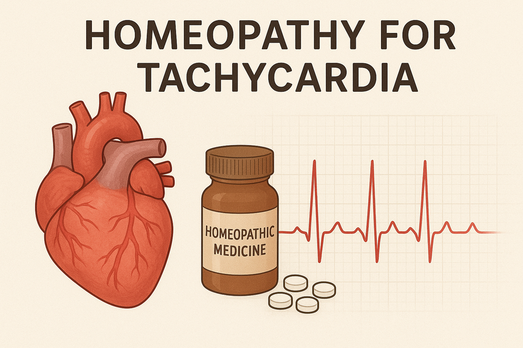
Homeopathy lacks robust evidence for tachycardia and should not replace conventional evaluation or urgent treatment. Some practitioners use remedies like Natrum muriaticum or Arsenicum album for palpitations anecdotally, but randomized controlled trials are insufficient to support clinical recommendations. Discuss any complementary therapies with your cardiologist to avoid interactions or delays in necessary interventions.
Putting It Together: Practical Steps You Can Take
If you feel a sudden rapid heartbeat, check your pulse and note the rate, regularity, associated symptoms, and duration. Try a vagal maneuver if the episode is brief and you have no chest pain or dizziness. Seek emergency care for syncope, chest pain, severe dyspnea, or sustained rates >150 bpm with instability. For recurrent, nonemergent palpitations, arrange prompt outpatient evaluation: ECG, ambulatory monitoring, basic labs (electrolytes, thyroid function), and echocardiography as indicated.
Long-term, focus on identifying triggers—caffeine, stimulants, dehydration, sleep deprivation, and unmanaged anxiety are common culprits—and discuss tailored medical and procedural options with your electrophysiologist. Combining evidence-based conventional treatments with selected lifestyle and complementary therapies gives you the best chance to control symptoms and lower risk.
Key Takeaways:
- Definition: Tachycardia is a resting heart rate >100 beats per minute and may be physiological (exercise, stress) or pathological (cardiac arrhythmia).
- Common causes: fever, anemia, hyperthyroidism, dehydration/hypovolemia, stimulants (caffeine, drugs), ischemia, electrolyte imbalances, and structural heart disease.
- Main types: sinus tachycardia, supraventricular tachycardia (including paroxysmal), atrial tachycardia, junctional tachycardia, ventricular tachycardia (monomorphic, polymorphic, and pulseless), and POTS.
- Symptoms: palpitations, rapid pulse, dizziness, shortness of breath, chest pain, syncope, and fatigue.
- Emergency signs: unstable blood pressure, severe chest pain, loss of consciousness, or pulseless ventricular tachycardia require immediate CPR/defibrillation and ACLS.
- Medical treatments: rate control (beta-blockers, calcium-channel blockers), antiarrhythmics, electrical cardioversion, catheter ablation, and treatment of underlying causes.
- Holistic approaches: lifestyle changes (remove stimulants, hydrate, correct electrolytes, regular exercise, weight management, and stress-reduction/breathing techniques); acupuncture may help some symptoms, but evidence is limited. homeopathy lacks robust evidence and should not replace medical care—consult a cardiologist before using alternatives.
The Biophysics of a Racing Heart
How Heart Rate is Measured
On an ECG, the most direct measurement of heart rate comes from the R‑R intervals: you measure the distance between successive R waves and convert that interval to beats per minute. The standard 12-lead ECG records at 25 mm/s and 10 mm/mV, so the quick bedside method—counting large boxes between R waves and using 300 for the number of large boxes—gives an immediate estimate; for more precision, you divide 1500 by the number of small 1 mm boxes between R waves. Holter monitors sample continuously (usually 128–1024 Hz for clinical devices) and deliver averaged rates across minutes or hourly windows, revealing patterns you would miss on a single 10‑second 12‑lead: A 24-hour Holter monitor may reveal that brief episodes of paroxysmal supraventricular tachycardia (PSVT) peak at 180–220 bpm during palpitations, whereas your resting ECG shows a rate of 72 bpm.
You can also measure rate with pulse palpation, pulse oximetry, chest‑strap ECGs, and photoplethysmography (PPG) in wrist wearables. Pulse palpation and simple portable monitors can make mistakes when measuring heart rhythms that are irregular, like in atrial fibrillation, which can cause the pulse to vary a lot from minute to minute. Consumer smartwatches that use PPG usually show heart rate within ±5–10 bpm when you’re steady, but they can be less accurate during quick movements or very fast heart rates; chest-strap monitors that track electrical activity are much more accurate, similar to clinical ECGs, and will match the R-R interval calculations used by Holter systems.
Heart rate variability (HRV) metrics add a layer of biophysical insight beyond mean beats per minute: SDNN, RMSSD, and frequency‑domain values (LF, HF, and LF/HF ratio) quantify autonomic modulation of your sinus node. Low SDNN values (for example, less than 50 ms over 24 hours) are often found with ongoing fast heart rates and a strong sympathetic nervous system response seen in conditions like reflex tachycardia or anxiety-related Implantable loop recorders give the longest continuous view (years) and are often used when your symptomatic episodes are rare but severe—for example, intermittent polymorphic ventricular tachycardia that occurs only during exertion and evades short monitoring periods.
Normal vs. Excessive Heart Rates
Clinically you classify tachycardia as a sustained heart rate above 100 beats per minute at rest; the range you consider “normal” depends on context—60–100 bpm is the accepted adult resting range, while athletes frequently rest below 60 bpm due to increased vagal tone. Sinus tachycardia typically sits between 100 and 140 bpm during physiologic states like fever or exercise, but rates above 140–150 bpm should prompt evaluation for supraventricular tachycardia (SVT) or atrial tachycardia. Paroxysmal SVT and junctional tachycardia commonly produce rates in the 150–250 bpm window, with sudden onset and termination distinguishing them from gradual sinus tachycardia linked to stressors or medication effects.
Ventricular tachycardia (VT) occupies a different clinical threshold: sustained VT is generally defined as a ventricular rate over 100–120 bpm lasting longer than 30 seconds or causing hemodynamic compromise, and many sustained VTs are in the 120–250 bpm range. Pulseless ventricular tachycardia—when your monitor shows rapid ventricular complexes without a palpable pulse—is a cardiac arrest rhythm requiring immediate defibrillation per ACLS algorithms; you should treat it as an emergency rather than a rate abnormality. In orthostatic conditions such as postural orthostatic tachycardia syndrome (POTS), you may see a rise of ≥30 bpm within 10 minutes of standing (≥40 bpm in adolescents), often without absolute rates exceeding 120 bpm, yet symptom burden can be as severe as higher‑rate arrhythmias.
Context matters: a 110 bpm sinus tachycardia during moderate exercise is physiologic, whereas 110 bpm at rest in a person with signs of ischemia or syncope is pathologic. You should factor in symptoms, hemodynamics, and rhythm origin—atrial versus ventricular—when interpreting whether a rate is excessive for a given patient. Ambulatory monitoring often helps clear up confusion: a study found that 48-hour Holter monitoring changed treatment for over 25% of patients with unexplained palpitations by detecting short episodes of non-sustained VT or sustained SVT that didn’t cause symptoms during their clinic visits.
Additional nuance comes from age and comorbidities: infants and children have higher normal resting rates (newborns 100–160 bpm, toddlers 90–150 bpm), and medications (beta‑agonists, anticholinergics, and stimulants) can raise your baseline by 10–40 bpm. Problems with electrolytes, like low potassium and magnesium levels, can make you more likely to have certain types of VT, even at heart rates that are usually considered normal for adults. Additionally, having heart problems or reduced blood flow makes it easier for a heart rate to become dangerous, so it’s important to consider these numbers along with the
The Diverse Faces of Tachycardia
Sinus Tachycardia: The Body’s Response to Stress
Sinus tachycardia is often recognized as the simplest electrical pattern on an ECG, characterized by a normal P wave preceding every QRS complex and a heart rate exceeding 100 beats per minute; however, its causes can vary from benign physiological responses to serious systemic illnesses. Exercise commonly pushes your sinus rate well past 100 bpm; fever raises metabolic demand so that each 1°C increase can raise heart rate by roughly 10–20 bpm. Dehydration and blood loss reduce preload and trigger compensatory sinus tachycardia; similarly, hyperthyroidism can sustain resting rates in the 100–140 bpm range, and stimulants such as 200–400 mg of daily caffeine or short-acting inhaled beta-agonists frequently provoke episodes you feel as palpitations.
Your clinical approach should focus on context: an otherwise healthy 25-year-old with a transient HR of 110–120 bpm after a sprint differs from a 70-year-old with fever, hypotension, and a sinus rate of 130 bpm during sepsis. Holter monitoring helps quantify burden—for example, inappropriate sinus tachycardia (IST) is considered when resting rates exceed 100 bpm with average 24-hour rates often above 90–95 bpm and symptomatic palpitations that are disproportionate to physiological triggers. Lab tests (like TSH, CBC, and electrolytes), checking blood pressure when standing up, and reviewing medications (like decon
Your treatment options reflect cause and severity: simple measures like oral rehydration, reducing caffeine intake, and treating fever or anemia will normalize many sinus tachycardias. If symptoms continue even after treating reversible factors, medication might be necessary—low-dose beta-blockers like metoprolol or atenolol can help lower high heart rates, while ivabradine (a specific type of heart medication) has been shown to help with symptoms. Gradual aerobic conditioning also lowers resting heart rate over weeks; one randomized rehab study showed a 7–10 bpm reduction in resting HR after 8–12 weeks of structured training.
Atrial Tachycardia: The Heart’s Electric Show
You may notice atrial tachycardia as sudden-onset palpitations with rates typically between 100 and 250 bpm originating from a single atrial focus, distinct from sinus rhythm by abnormal P-wave morphology or axis on ECG. Paroxysmal atrial tachycardia happens in episodes and can be triggered by stress hormones, heart damage like scarring from surgery, or digitalis toxicity; for instance, after coronary artery bypass grafting (CABG), up to 30% of patients may experience atrial tachyarrhythmias. Symptom severity ranges from mild palpitations to presyncope or syncope when ventricular response is rapid.
Your diagnostic tools include a standard 12-lead ECG, which may show P′ waves before each QRS with a consistent RP or PR pattern, and ambulatory monitoring to catch short episodes—a 48–72 hour Holter monitor improves diagnosis, while longer patch monitors or implantable loop recorders assist with rare episodes. Electrophysiology (EP) study pinpoints focal vs. reentrant mechanisms: focal atrial tachycardia produces centrifugal activation from one site (e.g., crista terminalis, pulmonary vein ostia), whereas macroreentrant circuits behave differently on mapping. Adenosine often helps identify where the heart’s fast beats are coming from by temporarily stopping signals to the AV node, which shows
Your management depends on stability and mechanism: vagal maneuvers and adenosine can terminate some paroxysms; intravenous procainamide or amiodarone is frequently used for hemodynamically stable episodes. For recurrent symptomatic focal AT that resists medication, catheter ablation offers definitive therapy with success rates of 70–90% depending on location and complexity. Decisions about long-term anticoagulation are based on the risk of stroke when atrial tachycardia occurs alongside clinical atrial fibrillation or
Additional nuance appears in differentiating focal atrial tachycardia from multifocal atrial tachycardia (MAT): MAT shows three or more distinct P-wave morphologies and irregular R‑R intervals, commonly associated with COPD and hypoxemia, whereas focal AT is usually regular and monomorphic. Treatment differs—fixing low oxygen levels and lung problems helps MAT, while focal AT might need specific procedures or medications; understanding this difference helps prevent giving unnecessary medications to patients whose heart rhythm issues are caused by lung disease.
Ventricular Tachycardia: When the Heart Goes Rogue
You confront ventricular tachycardia (VT) as one of the most threatening arrhythmias—sustained VT usually presents with rates of 120–250 bpm and arises from the ventricles, not the atria, causing wide QRS complexes and potentially rapid hemodynamic compromise. Ischemic scar after myocardial infarction is the single most common substrate, producing monomorphic VT from a stable reentrant circuit within scar tissue; polymorphic VT, including torsades de pointes, is associated with prolonged QT and can degenerate to ventricular fibrillation (VF) and sudden cardiac death. Electrolyte disturbances (K+ 5 mEq/L, Mg2+ <1.8 mg/dL) and QT‑prolonging drugs dramatically increase risk.
Your immediate treatment depends on the patient’s condition: if the patient has unstable VT with low blood pressure, chest pain, or fainting, you need to perform synchronized cardioversion right away; if the patient has pulseless VT, follow the VF protocol. Stable monomorphic VT can be treated with antiarrhythmics like IV procainamide or amiodarone, but it’s crucial to find and fix any underlying issues like lack of In the intensive setting, temporary measures like overdrive pacing or sedation may be used while you plan definitive therapy.
Your long-term plan aims to stop the heart rhythm problems from coming back and to prevent sudden death: implantable cardioverter-defibrillators (ICDs) are recommended for people who have survived VT/VF or have ongoing VT with serious left ventricular dysfunction. Catheter ablation that targets reentry circuits related to scar tissue can cut down the number of ICD shocks by 50–70% in skilled centers; for polymorphic VT linked to QT issues, the first steps are giving magnesium sulfate and stopping any problematic medications, along with pacing to Prognosis varies: post-MI patients with LVEF <35% have markedly higher VT recurrence and mortality, guiding more aggressive device and ablation therapy.
Further practical detail concerns pulseless VT and ACLS specifics: after immediate defibrillation and CPR, administration of epinephrine 1 mg every 3–5 minutes is recommended, and amiodarone 300 mg IV bolus (followed by 150 mg if needed) is the antiarrhythmic of choice in refractory cases. Rapid identification of reversible causes—the Hs and Ts (hypovolemia, hypoxia, hydrogen ion/acidosis, hypo-/hyperkalemia, hypothermia; toxins, tamponade, tension pneumothorax, thrombosis)—directly informs both resuscitation and post‑resuscitation planning.
Postural Orthostatic Tachycardia Syndrome: The Dizzying Twist
You will recognize POTS by a reproducible orthostatic heart rate rise: adults meet diagnostic criteria with an increase of ≥30 bpm within 10 minutes of standing (or ≥40 bpm for adolescents) without orthostatic hypotension. Symptoms include lightheadedness, palpitations, presyncope, chronic fatigue, and cognitive complaints often labeled “brain fog.” More women aged 15–50 are affected, and many patients say their symptoms started after a viral illness, like Epstein–Barr or COVID-19, which suggests issues with the autonomic nervous system or small-fiber nerves.
To start your evaluation, you should check for changes in blood pressure and heart rate with active stand tests and tilt-table testing if the diagnosis is not clear; 24-hour Holter monitoring often shows increased sympathetic activity, with average heart rates 10–20 beats per minute higher than normal. You can start managing the condition with simple steps:increase your daily salt intake to 3–5 grams, drink 2–3 liters of fluids, wear waist-high compression garments, and follow a gradual exercise program that begins with cycling while lying down and moves to upright training over 3–6 months—studies have shown that this approach can help improve your ability to stand and reduce symptoms when done with
Your pharmacologic toolkit includes fludrocortisone to expand plasma volume, midodrine for venoconstriction, low-dose propranolol or ivabradine to blunt heart rate surges, and selective use of SSRIs or SNRIs when comorbid autonomic instability and mood symptoms coexist. How well patients respond can vary: a 2014 study found that about 60–70% of patients felt better with a mix of non-drug and drug treatments, but it’s important to adjust treatments for each person and keep track of their progress because how well they handle
Additional tests are needed to tell the difference between POTS, orthostatic hypotension, and anxiety disorders; tilt-table testing helps understand how the body works, while autonomic reflex testing and If you have long-lasting symptoms after an infection or serious difficulties with daily activities, getting help from a team of specialists—like heart doctors, brain doctors, physical therapists, and sometimes immune system experts—can lead to better results
Exploring the Causes: What Triggers Your Heart Racing?
Physiological Causes: From Exercise to Fever
Your heart rate rises predictably with physical exertion because cardiac output must increase to meet skeletal muscle oxygen demands; during moderate-intensity exercise your pulse commonly climbs into the 120–160 bpm range depending on age and fitness, and elite athletes may transiently exceed 180 bpm during maximal efforts. A fever elevates your metabolic rate, and for every 1°C increase in body temperature, your heart rate typically increases by about 10 beats per minute, so a 39°C fever can add roughly 20–30 bpm to your resting rate. Dehydration reduces circulating volume, prompting a compensatory tachycardia to preserve perfusion—athletes, outdoor workers, and older adults who lose 1–2% of body weight through sweating can already notice palpitations as blood pressure dips.
Your autonomic balance shifts toward sympathetic dominance with many everyday physiological stressors: caffeine and nicotine stimulate beta-adrenergic receptors, certain cold medications with pseudoephedrine provoke reflex tachycardia, and even a heavy meal can temporarily increase heart rate as digestion raises metabolic demand. When you change positions, your body reacts in specific ways—postural orthostatic tachycardia syndrome (POTS) causes your heart rate to increase by 30 beats per minute or more (or to over 120 beats per minute) within ten minutes of standing up, without a drop Pregnancy creates a chronic high-output state; blood volume increases by 30–50% and resting heart rate commonly rises by 10–20 bpm, so palpitations during gestation are frequently physiological but still warrant evaluation if severe or accompanied by other symptoms.
Circumstantial factors interact: combining fever, dehydration, and stimulant intake amplifies tachycardia more than any single factor alone, which explains why you might feel your heart racing far more on a hot, caffeinated afternoon than during a calm morning run. Acute hypoxia—whether from high-altitude exposure, COPD exacerbation, or pulmonary embolism—provokes sympathetic surges and rapid breathing that drive heart rate upward as the body tries to maintain oxygen delivery. When doctors evaluate an episode, they look for patterns in your body’s responses—like when you exercised, the temperature, how much fluid you lost, if you had any stimulants, and your body position—to tell the difference between normal sinus tachy
Psychological Factors: Stress and Anxiety as Catalysts
Acute psychological stress triggers the body’s stress response, leading to the release of adrenaline and noradrenaline, which speeds up the heart rate; during panic attacks, your heart rate can jump to 120–180 bpm in just a few minutes, often causing chest tightness, shortness of breath, and a sense of impending doom. Chronic anxiety disorders keep baseline sympathetic tone elevated, producing persistent sinus tachycardia and frequent palpitations that may mimic intermittent supraventricular tachycardia; studies show individuals with generalized anxiety report palpitations at significantly higher rates than control groups. Hyperventilation during anxiety episodes changes carbon dioxide levels and can provoke lightheadedness and perceived racing, which then feeds back into more anxiety and sustained tachycardia.
Situational stressors—public speaking, acute grief, high-pressure work deadlines—trigger reproducible heart rate responses; laboratory stress tests document 20–40 bpm increases in healthy volunteers exposed to timed cognitive tasks. Post-traumatic stress disorder and panic disorder demonstrate particularly strong autonomic dysregulation, with elevated nocturnal heart rates and decreased heart rate variability, markers linked to higher cardiovascular risk over time. If you notice palpitations that consistently correspond to social, occupational, or emotional triggers, targeted interventions such as breathing retraining, cognitive behavioral therapy, or biofeedback frequently reduce both subjective distress and measurable heart rate spikes.
Certain personality and behavioral patterns compound the effect: high trait anxiety, avoidance behaviors that reduce physical conditioning, and reliance on stimulants to cope all make you more susceptible to recurring episodes. Vagal maneuvers—bearing down, splashing cold water on the face—may abort some supraventricular episodes but are less effective against anxiety-driven sinus tachycardia, where addressing the psychological driver is more successful for long-term control. Exposure-based therapies and mindfulness programs have shown reductions in panic frequency and average resting heart rate in randomized trials, supporting a mind–body approach when emotional triggers dominate the clinical picture.
- Acute panic attacks often present with HR surges into the 120–180 bpm range and intense subjective fear.
- Chronic anxiety can produce persistent resting sinus tachycardia and reduced heart rate variability on Holter monitoring.
- POTS and anxiety frequently coexist, complicating diagnosis and requiring orthostatic testing.
- Any high-intensity emotional event or repeated stress exposure can precipitate recurrent palpitations and should be evaluated in the context of your overall health.
Worsening sleep quality and nighttime hyperarousal commonly amplify daytime palpitations: fragmented sleep elevates sympathetic tone, and shift work or insomnia can prevent the nocturnal heart rate dip that normally restores autonomic balance, leaving you more prone to daytime tachycardia. Psychological comorbidity also alters symptom perception—patients with anxiety often report palpitations at lower absolute heart rates than those without, so symptom burden may not correlate directly with ECG findings. Integrated care that combines psychiatric assessment, behavioral therapy, and, when appropriate, short-term pharmacologic support yields the best outcomes in reducing both the subjective experience of racing and objective rate abnormalities.
- Breathing exercises and paced respiration improve CO₂ regulation and blunt hyperventilation-induced palpitations.
- CBT and exposure therapy reduce panic frequency and lower resting sympathetic activity over months of treatment.
- Biofeedback and heart rate variability training can increase vagal tone and reduce palpitations in controlled trials.
- Any persistent or worsening symptom should prompt medical evaluation to exclude overlapping cardiac or endocrine causes.
Medical Conditions: The Role of Thyroid Disorders and Anemia
Hyperthyroidism accelerates metabolic processes and increases beta-adrenergic receptor sensitivity, so you often experience resting sinus tachycardia, exercise intolerance, and tremor; typical lab findings include suppressed TSH (often <0.1 mIU/L) with elevated free T4 and T3. Older adults with thyrotoxicosis have a higher incidence of atrial fibrillation—reported rates range from 10% to 25% depending on age and comorbidities—so a new-onset irregularly irregular pulse in someone with heat intolerance, weight loss, and tremor should trigger thyroid testing. Subclinical hyperthyroidism can also produce palpitations and subtle heart rate increases that affect quality of life, even when T4/T3 are within reference ranges but TSH is low.
Anemia decreases arterial oxygen content, compelling the cardiovascular system to raise heart rate and stroke volume to preserve oxygen delivery; clinically significant tachycardia commonly emerges when hemoglobin falls below about 10 g/dL, though symptomatic palpitations can occur at higher levels depending on baseline fitness and comorbid lung or cardiac disease. Iron-deficiency anemia is a frequent, treatable cause—women of reproductive age, patients with gastrointestinal blood loss, and chronic kidney disease populations are particularly at risk—and a complete blood count plus ferritin level will clarify the diagnosis. In severe or rapidly developing anemia, you may see tachycardia accompanied by orthostatic symptoms, hypotension, and signs of high-output heart failure due to chronically increased cardiac workload.
Other health issues that can cause a fast heartbeat include sepsis, where heart rates often go above 90–100 bpm as part of the body’s response, and lung problems—pulmonary embolism usually causes a sudden fast heartbeat that is often much greater than what you’d expect from chest When you present with unexplained palpitations, clinicians screen for endocrine and hematologic abnormalities because correcting hyperthyroidism or replenishing iron frequently normalizes heart rate without the need for long-term rate-control medications. Diagnostic workups typically include TSH, free T4, CBC with indices, ferritin, and, when indicated, ECG and ambulatory monitoring to document rhythm during symptomatic episodes.
Some clinical vignettes illustrate the overlap: a 68-year-old man with atrial fibrillation whose only systemic complaint was weight loss and heat intolerance was found to have TSH < 0.01 and underwent treatment that restored sinus rhythm after euthyroid status; a 35-year-old woman with heavy menstrual bleeding and hemoglobin of 8.2 g/dL reported daily palpitations that resolved within weeks of iron repletion. These examples show how addressing the underlying thyroid or hematologic disorder often removes the trigger for your tachycardia and reduces reliance on symptomatic therapies.
Recognizing the Red Flags: Symptoms that Signal Trouble
Common Symptoms: From Palpitations to Shortness of Breath
Palpitations usually present as a sudden awareness of your heartbeat—described by many patients as pounding, fluttering, or a rapid thudding in the chest—and often signal a quick rhythm such as sinus tachycardia or atrial tachycardia. You may notice palpitations triggered by caffeine, nicotine, fever (for example a temperature of 38.5°C raising your resting rate above 100 bpm), dehydration, or anxiety; in other cases, they appear abruptly with episodes of paroxysmal supraventricular tachycardia (PSVT), where rates commonly reach 150–250 beats per minute. Pay attention to the pattern: brief, self-limited bursts that stop within seconds to minutes are more typical of benign SVT, while sustained, repetitive episodes or palpitations accompanied by lightheadedness suggest a higher-risk arrhythmia such as ventricular tachycardia (VT).
Shortness of breath during tachycardia often reflects reduced cardiac output and impaired ventricular filling, particularly when rates exceed 120–140 bpm and diastolic filling time shortens. You might feel breathless with minimal exertion or even at rest; pulmonary congestion and orthopnea develop when the rapid rate precipitates acute heart failure, a scenario seen in older patients or those with prior myocardial infarction. In postural orthostatic tachycardia syndrome (POTS), standing causes a heart rate rise of ≥30 bpm (or >120 bpm) within 10 minutes, and you experience dizziness, cognitive fog, and breathlessness that improve when reclining.
Red flags such as dizziness, near-syncope, and syncope signal cerebral hypoperfusion due to decreased stroke volume during tachyarrhythmia. NNon-sustained VT, which lasts less than 30 seconds, can cause brief lightheadedness, while sustained VT lasting more than 30 seconds or fast atrial fibrillation often leads to fainting or collapsing; in serious cases of VT, heart rates typically range from 120 to 250 bpm. Chest pain that comes with a fast heartbeat should raise concerns about reduced blood flow to the heart—common signs include a pressure-like pain that spreads to the arm or jaw, especially in patients with heart disease where a fast heartbeat demands more oxygen than the heart can supply.
When to Seek Immediate Help for Tachycardia: Identifying Emergency Situations
Loss of consciousness, ongoing chest pain, severe breathlessness, or sudden confusion during a rapid heartbeat are emergency signs that require immediate evaluation—call emergency services without delay. Hemodynamic instability means low blood pressure (systolic blood pressure <90 mmHg), ongoing dizziness, or signs of shock, which often shows that the fast heartbeat (like sustained monomorphic VT at 150–200 bpm) is affecting the heart’s ability to pump. Patients with structural heart disease, recent myocardial infarction, or known cardiomyopathy should lower their threshold for seeking help because even moderate increases in rate can precipitate decompensation.
If there is no pulse when someone has a fast, unresponsive heartbeat, it means they might have pulseless ventricular tachycardia or ventricular fibrillation, which requires immediate CPR and defibrillation following Survival from VF/pulseless VT declines sharply with time; studies estimate a 7–10% decrease in survival for each minute without defibrillation, so bystander CPR and early use of an automated external defibrillator (AED) dramatically affect outcome. If you witness someone collapse with a rapid abnormal rhythm and they are unresponsive and not breathing normally, activate emergency services, start chest compressions, and apply an AED as soon as possible.
Sustained rapid rhythms that produce recurrent presyncope, ongoing ischemic chest pain, or progressive shortness of breath despite rest also require urgent care even if blood pressure remains borderline. Emergency departments will usually do a quick 12-lead ECG, monitor your heart, and use bedside echocardiography to check what type of arrhythmia you have (like atrial tachycardia or VT), how well your heart is working, If you have an implanted device (pacemaker or ICD) and experience shocks, rapid deterioration, or persistent symptoms after a shock, head to the nearest emergency room because device interrogation and possible reprogramming or antiarrhythmic therapy may be needed.
Immediate self-actions can change outcomes: avoid driving yourself to the hospital if you feel faint; lie flat and elevate your legs if lightheaded while awaiting help to improve cerebral perfusion; if chest pain is present and you are not allergic and have been advised previously, chew aspirin while emergency services are en route. Bystanders should prioritize calling emergency services, initiating CPR for unresponsiveness with no pulse, and using an AED for any suspected pulseless VT or VF—early intervention is the single most impactful step in survival from life-threatening tachyarrhythmias.
Beyond Conventional Medicine: Exploring Holistic Approaches
Natural Remedies: Dietary and Lifestyle Adjustments
You can reduce episodic and baseline tachycardia risk by correcting common dietary drivers: limit caffeine to about 200–300 mg daily (roughly 2–3 cups of brewed coffee), cut back on alcohol to the recommended limits (up to 1 drink per day for women, up to 2 for men), and aim for a DASH-style or Mediterranean pattern rich in vegetables, legumes, whole grains and oily fish. Electrolyte balance is relevant for rhythm stability—prioritize dietary magnesium and potassium from spinach, nuts, avocados, bananas and beans rather than high-dose supplements unless prescribed; typical supplemental magnesium dosages used for arrhythmia support range from 200 to 400 mg/day, but you should coordinate that with your clinician if you take diuretics or have kidney disease. Reducing sodium to under 2,300 mg/day can also help if you have concurrent hypertension, since high blood pressure and volume shifts commonly provoke reflex tachycardia and atrial arrhythmias.
You can change your daily habits to lower sympathetic triggers: aim for 150 minutes of moderate aerobic activity weekly (brisk walking, cycling), which typically corresponds to 50–70% of your estimated maximum heart rate (220 minus your age), and add two strength sessions. Manage body weight toward a BMI in the 18.5–24.9 range if feasible; excess adiposity increases circulating catecholamines and inflammatory cytokines that raise resting heart rate and the likelihood of atrial and sinus tachycardia. Monitor hydration closely—losing as little as 2% of body weight from fluid loss can increase heart rate by 10–20 beats per minute, especially relevant if you have postural orthostatic tachycardia syndrome (POTS) or are endurance-training.
You should check your medications and over-the-counter products for stimulants like pseudoephedrine, nasal decongestants, some thyroid supplements, high-dose vitamin B12/energy shots, and certain herbal stimulants (like ma huang/ephedra and high-dose green). Keep a log of episodes related to foods, drinks, and activities; wearable devices that record heart rates over 24 to 72 hours can reveal patterns (for example, episodes clustered after consuming late-afternoon coffee or during nights of poor sleep). Coordinate any dietary changes and supplement starts with your cardiology or primary care team, particularly if you take antiarrhythmics, beta-blockers, anticoagulants, or other cardiac medications.
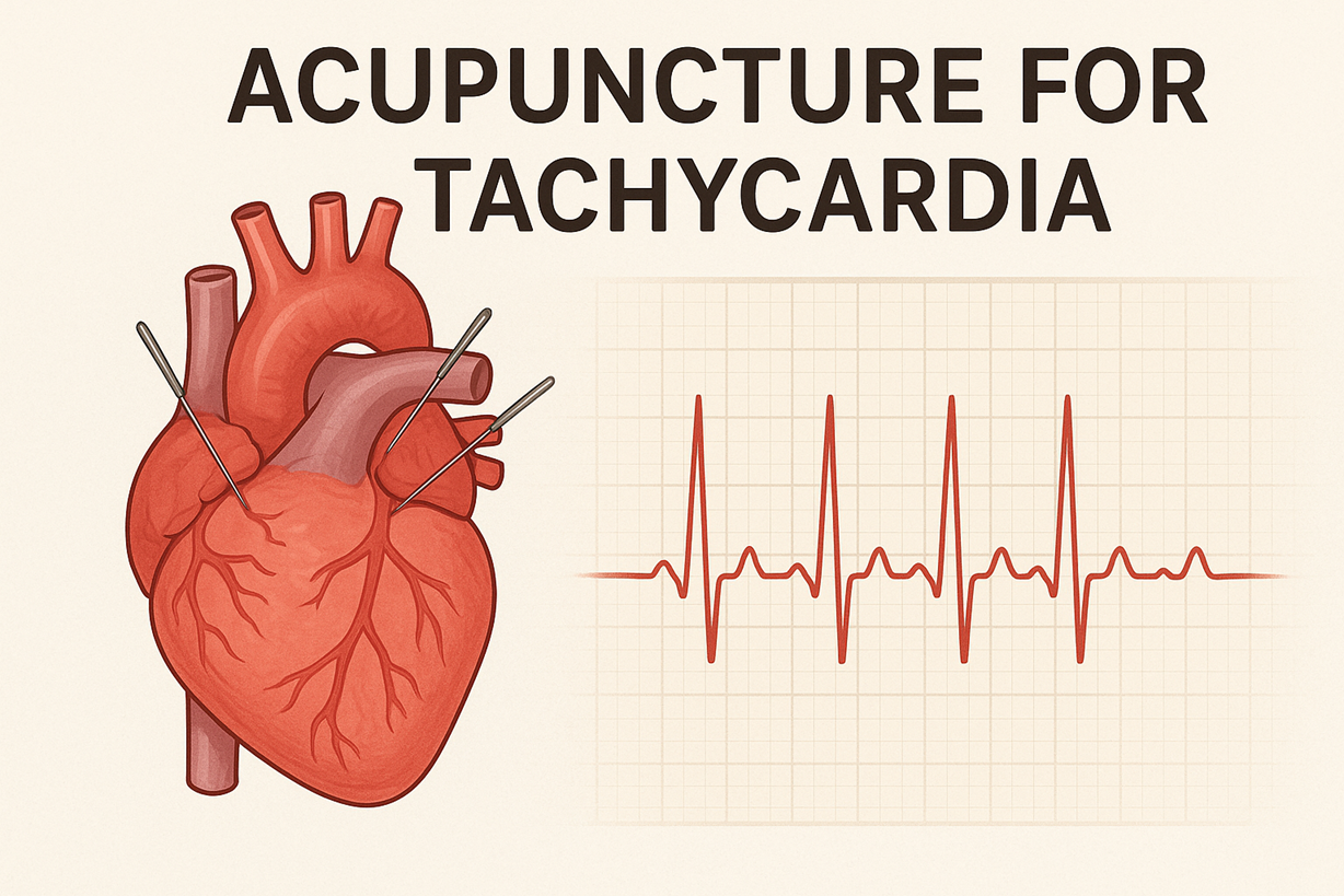
Acupuncture and Homeopathy: Alternative Paths to Relief Tachycardia
You can try acupuncture to help with balance in your body and to reduce palpitations; practitioners often use specific points like PC6 (Neiguan), HT7 (Shenmen), and ST36 to help with vagal tone and anxiety. Clinical reports and small studies have shown that acupuncture can help reduce palpitations, anxiety, and changes in heart rate, but the results can vary depending on the technique used and the practitioner’s training. Expect treatment sessions to last between 20 and 40 minutes using clean, single-use needles; sometimes, low-frequency electroacupuncture (2–10 Hz) is added for stronger effects on the nervous system and is generally well accepted
You can see a trained homeopathic practitioner to help find remedies that match your symptoms if you prefer treatments with very diluted substances; common homeopathic remedies for palpitations include Aconitum napellus, Arsenicum album, and sometimes Crataegus preparations (hawthorn), which are used as herbal heart tonics instead of homeopathic dilutions. There is not much strong evidence for homeopathy in treating tachycardia: some reports show that a few patients feel better, but there aren’t many well-controlled studies, and the results vary widely. Make sure you disclose all prescription drugs to any alternative provider, because herbal cardiotonics like hawthorn can interact with digoxin and blood-pressure medications, and overlapping therapies may alter your clinical monitoring needs.
Many clinics combine acupuncture with breathing exercises or mindfulness to enhance the benefits; doctors usually suggest keeping up with regular heart check-ups using ECG or Holter monitoring while trying alternative therapies, and some programs track heart rate variability (HRV) or symptom scores before and after treatment to show changes over 6–12 weeks.
Mindfulness and Relaxation Techniques: Calming the Heart
You can reduce sudden increases in heart activity and their frequency by using proven breathing and relaxation techniques. Breathing at a steady pace of about 6 breaths per minute for 10 to 20 minutes twice a day can help increase heart rate variability (HRV) and may lower your resting heart rate, with some studies indicating a decrease of several beats per minute. Biofeedback programs for heart rate variability help you learn your personal rhythm for breathing and practice breathing in and out at a steady pace with visual or sound signals; these programs usually last 4 to 8 weeks and involve daily 15- to 20 minute sessions at home along with weekly coaching, and small studies have shown they can lead to fewer heart palpitations in patients with paroxysmal tachy
You can enroll in an 8-week Mindfulness-Based Stress Reduction (MBSR) course or use targeted practices, such as progressive muscle relaxation, guided imagery, and body scans, to reduce baseline sympathetic tone. MBSR courses typically include weekly group sessions lasting 2 to 2.5 hours and require 30 to 45 minutes of daily practice at home; research shows that these courses can help reduce anxiety, improve sleep, and slightly lower resting heart rate and blood pressure, which together can decrease triggers for irregular heartbeats. Adding gentle exercises like restorative yoga or tai chi two to three times a week provides both aerobic benefits and helps calm the nervous system, which can be especially useful for patients with sinus tachycardia or POTS who can handle light, low-impact exercise.
You can pair mindfulness with technology-based supports: smartphone apps offering guided paced-breathing, wearable HRV coaching, and short (5–12 minute) “micro-practices” are practical for acute palpitations—use them at the onset of symptoms to lower anxiety and blunt sympathetic escalation. Track your episodes alongside your mindfulness practice; some patients notice a drop in episode intensity within 2–6 weeks of consistent practice, and measurable HRV improvements commonly appear after 4–8 weeks of daily training. Coordinate these practices with your cardiology care, especially if you experience syncope or sustained ventricular arrhythmias or are on antiarrhythmic medications.
More detailed logistical tips: start with short, reproducible protocols (for example, 10 minutes of 6-breaths-per-minute breathing in the morning and evening) and add biofeedback or group MBSR if you need structured support; clinicians often recommend keeping a simple symptom-and-practice diary to correlate practice adherence with changes in palpitation frequency and intensity over a 6–12 week period.
Building a Personalized Action Plan: Your Road to Recovery
Monitoring Your Heart Health: Tools and Techniques
Home and clinical monitoring together give you the best chance of correlating symptoms with rhythm abnormalities. Ambulatory ECG options include a 24–48 hour Holter, which catches frequent daily events; a 2–4 week patch monitor for intermittent episodes; and an event recorder or external loop monitor that you can trigger when symptoms start. Implantable loop recorders (ILRs) can record continuously for up to three years and are ideal if you have infrequent but disabling palpitations or syncope. Wearable devices like smartwatches and chest straps that can do While ECGs are effective at detecting atrial fibrillation and fast heart rhythms in many individuals, their accuracy can vary significantly. research shows that smartwatch ECGs find AF about 90% of the time in people with symptoms, but there are more false alarms if you have occasional heartbeats or if you’re
Quantifying episodes requires more than a strip of rhythm data: you should log context—activity, caffeine/alcohol intake, medications, stress levels, posture changes, and associated symptoms (dizziness, chest pain, breathlessness). Orthostatic measurements matter if orthostatic intolerance or POTS is suspected: measure heart rate and blood pressure supine and at 1, 3, and 10 minutes after standing; an increase of ≥30 bpm in adults (≥40 bpm in adolescents) within 10 minutes supports a POTS diagnosis. For suspected ventricular tachycardia, document rate and duration—sustained VT is usually defined as >100–120 bpm lasting >30 seconds or causing hemodynamic compromise. Combining symptom diaries with objective monitors helps your clinician distinguish sinus tachycardia, supraventricular tachycardia, and ventricular arrhythmias and tailor therapy accordingly.
Learn and practice simple vagal maneuvers and vital-sign techniques so you can act safely when an episode begins. The modified Valsalva maneuver (15 seconds of strain at ~40 mmHg using a mouthpiece or syringe, then rapid supine reposition and leg raise) restores sinus rhythm in a significant proportion of AV nodal reentrant tachycardias—NEJM data showed higher cardioversion rates vs. standard Valsalva. Carotid sinus massage can be diagnostic and therapeutic but should only be done in a monitored setting after ruling out carotid stenosis or recent stroke. If you have an implanted device or are prescribed antiarrhythmic therapy, schedule periodic device interrogations and electrolyte checks (potassium 3.5–5.0 mmol/L, magnesium in the normal range) because electrolyte disturbances commonly precipitate arrhythmias.
Collaborating with Health Professionals about Your Tachycardia: Finding the Right Support
Start with a clinician who listens and takes your symptom timeline seriously: your primary care provider or an internal medicine physician can coordinate initial testing—ECG, basic labs including TSH and electrolytes, and ambulatory monitoring. If you have ongoing, noticeable, or unclear rapid heartbeats, you should see a cardiologist; if you need more detailed tests or treatments, an electrophysiologist (EP) will be important. Ask prospective specialists about their volume and outcomes:centers that perform more than 50–100 ablations per year typically report better procedural success and complication rates. When discussing specific procedure success rates, you can mention that AVNRT ablation has a success rate of over 95%, accessory pathway (WPW) ablation is about 90–95%, and paroxysmal AF ablation
Expect a step-by-step approach to diagnosis with the team:start with a baseline 12-lead ECG, then a transthoracic echocardiogram to check heart structure and ejection fraction (EF), do exercise stress testing if there’s a suspicion of exertional tachycardia, and use targeted imaging like If the outpatient tests don’t give clear answers and symptoms keep happening, an EP study can help identify where the arrhythmia begins and allow for catheter ablation at the same time.
Form a team of different specialists to address the causes of your fast heartbeat: consult an endocrinologist for hyperthyroidism, a neurologist for possible autonomic disorders like POTS, and a sleep specialist if you have obstructive sleep apnea, as it can increase the risk of heart rhythm problems and often improves with CPAP Mental health professionals and physiotherapists can help with anxiety-driven or deconditioning-related tachycardia through CBT and graded exercise programs; nutritionists can guide sodium and fluid strategies for POTS or weight-loss plans to reduce arrhythmia burden. Use telemedicine follow-ups for quick medication checks and device interrogations when appropriate, and ensure an emergency plan is in place: know when to call emergency services (syncope, severe chest pain, hypotension) versus contacting your clinic.
Lifestyle Changes: Sustainable Practices for Heart Rhythm Management
Dietary and fluid adjustments can have measurable effects on certain forms of tachycardia. If you have POTS, increasing daily fluid intake toward 2.5–3 liters and sodium intake to roughly 3–5 grams per day (under clinician guidance) often raises intravascular volume and reduces orthostatic tachycardia. For others, cutting back on stimulant exposures—caffeine to below ~200 mg/day (about two standard coffees), avoiding energy drinks with high-dose taurine and caffeine, and stopping recreational stimulants—reduces episodes of reflex and sinus tachycardia. Alcohol reduction matters too: binge drinking is a common trigger for atrial fibrillation (“holiday heart” phenomenon) and even moderate chronic intake increases AF risk; targeting no more than 1 drink/day for women and 2 for men aligns with arrhythmia-reduction strategies.
Exercise prescription should be individualized: aim for at least 150 minutes per week of moderate-intensity aerobic activity for general cardiovascular health, but if you have POTS or deconditioning, begin with recumbent or semi-recumbent training (recumbent cycling, rowing) and progress slowly—protocols that start with 20–30 minutes three times weekly and add 5–10 minutes every 1–2 weeks can restore autonomic balance and reduce resting tachycardia over months. Weight loss to a BMI in the healthy range reduces atrial dilation and AF risk; losing 10% body weight in obese patients has been associated with significant AF burden reduction in clinical studies. Addressing sleep apnea with CPAP often lowers nocturnal and daytime tachyarrhythmia frequency and should be prioritized when apnea is present.
Stress management and sleep hygiene produce durable benefits for rhythm control. Planned activities like cognitive behavioral therapy for anxiety, slow breathing (6 breaths per minute for 10 minutes each day), and mindfulness exercises help reduce stress and can lower episodes of fast heartbeats. Maintain regular sleep schedules targeting 7–9 hours per night and screen for contributory conditions like insomnia or restless legs. Stay away from over-the-counter products that stimulate the nervous system—like decongestants, certain weight-loss pills, and herbal stimulants such as ephedra—because even
Consider complementary therapies as adjuncts rather than replacements: low-risk options such as supervised acupuncture may reduce perceived palpitations and anxiety in some people, but high-quality evidence is limited and results are variable; always discuss supplements and alternative treatments with your cardiology team to avoid interactions with antiarrhythmic drugs or anticoagulants.
The Impact of Tachycardia on Daily Life: Balancing Activity and Rest
Understanding Limitations: Modifying Physical Activity
Assess your baseline using objective data: a resting heart rate above 100 beats per minute defines tachycardia in adults, but symptom thresholds vary by type. Arrange a Holter monitor or event recorder to capture episodes and an exercise stress test to map when your tachycardia is provoked. Use predicted maximum heart rate (220 − age) as a starting point, then work with your clinician to set a lower exercise ceiling—many patients are advised to keep exertion below 70–80% of predicted max or under an absolute heart rate limit (for example, <120–130 bpm) depending on whether you have sinus tachycardia, atrial tachycardia, POTS, or a history of ventricular tachycardia. Case example: a 48‑year‑old with atrial tachycardia discovered via Holter that exertional episodes began above 125 bpm and adjusted training to stay beneath that threshold with measurable symptom reduction.
Shift the type and structure of workouts rather than eliminating activity. Low‑impact aerobic options—recumbent cycling, swimming, elliptical, and rowing—reduce orthostatic stress for POTS and lower peak adrenergic surges for sinus tachycardia. Strength training should focus on higher repetitions with lighter loads and controlled breathing to avoid Valsalva maneuvers; aim for 2–3 resistance sessions weekly and 3–5 aerobic sessions of 20–40 minutes, progressing by about 10% per week from an initial 10–15 minute session if you tolerate it. Keep track of how hard you’re working by using a heart rate monitor and paying attention to how you feel (aim for a Borg score of 11–13); many people with sinus tachycardia find that their symptoms improve when they switch from intense workouts to moderate exercise, like steady walking, along with yoga
Factor in device status and medications when planning activity: if you have an ICD after ventricular tachycardia or are on antiarrhythmic therapy, avoid contact sports and activities with a high risk of trauma to the chest, and obtain cardiology clearance before returning to strenuous work. Practical safety steps include carrying a medical ID, programming your wearable to alarm at your individualized HR limit, and pausing exercise for symptoms such as dizziness, syncope, chest pain, or sustained palpitations. For POTS, adjunctive measures that enable increased activity include wearing compression stockings, increasing daily fluid intake to 2–3 liters, and consuming an additional 3–5 grams of salt per day as advised by your clinician; these simple modifications often allow for longer and safer exercise sessions without triggering symptomatic tachycardia.
Emotional Support: Coping Mechanisms for Living with Tachycardia
Expect a significant emotional component: studies report anxiety and depression in 30–40% of patients with arrhythmias and even higher rates in POTS. Look for therapies that address anxiety related to bodily sensations; cognitive behavioral therapy (CBT) specifically designed for heart patients can alleviate health concerns and reduce symptoms, as shown in many studies. Incorporate paced breathing (about six breaths per minute) or guided biofeedback sessions to boost heart rate variability and calm sympathetic overdrive; repeated practice of 10 minutes twice daily has measurable effects on vagal tone and subjective palpitations. Peer support networks and cardiac rehab programs tailored for arrhythmia patients improve quality of life and functional capacity; cardiac rehab can reduce readmission and help you regain confidence in graded activity.
Develop concrete in‑the‑moment strategies for episodes of rapid heart rate. Learn the modified Valsalva technique used in the REVERT trial, which increased termination of supraventricular tachycardia to 43% versus 17% for standard Valsalva; the maneuver involves a 15‑second strain followed immediately by a supine passive leg raise and facemask or syringe blow to maintain intrathoracic pressure. Vagal maneuvers such as carotid sinus massage should only be performed by trained providers; if you have a history of paroxysmal SVT, practice the modified Valsalva with your clinician so you can use it safely. Keep a small action plan accessible (medication list, when to call EMS, and the name of your electrophysiologist) and rehearse it so stress doesn’t impair decision-making during an episode.
Make workplace and social adjustments that preserve function and reduce isolation. Request flexible scheduling, regular hydration breaks, and temporary restrictions from solitary work at heights or around heavy machinery if you experience near‑syncope or unpredictable paroxysms; legal protections often allow reasonable accommodations. Train at least two coworkers in basic CPR and AED use if you carry an elevated risk of life‑threatening ventricular arrhythmia and keep an up‑to‑date emergency card and device ID in your wallet. Track triggers in a symptom diary or app—caffeine, alcohol, poor sleep, dehydration, and some over‑the‑counter decongestants are common precipitants—then use that dataset to make targeted lifestyle changes with your care team.
Use different types of help: combine follow-up care from heart specialists with therapy for mental health, supervised exercise from physical therapists or heart rehab, and support from patient groups like Dysautonomia International for POTS or national arrhythmia organizations for AF/VT. Pharmacologic options for comorbid anxiety or depression (for example, SSRIs) can be coordinated with your cardiologist to avoid drug interactions with antiarrhythmics. Establish measurable goals with your team—such as returning to a 30‑minute walk three times per week without palpitations—and use data from wearables and symptom logs to guide adjustments in both medical and behavioral therapies.
Summing up
On the whole, tachycardia refers to a fast heartbeat—generally an adult resting heart rate above about 100 beats per minute—and it can arise from many different mechanisms that affect your heart’s rate and rhythm. You may experience sinus tachycardia as a normal physiological response to exercise, fever, pain, anxiety or dehydration, or you may have arrhythmic forms such as atrial tachycardia, junctional tachycardia, paroxysmal supraventricular tachycardia, or the more dangerous ventricular tachycardias (including monomorphic and polymorphic varieties and pulseless ventricular tachycardia). Other recognized forms include reflex tachycardia and postural orthostatic tachycardia syndrome (POTS); underlying contributors often involve electrolyte imbalances, thyroid disease, anemia, structural heart disease, stimulants (caffeine, nicotine, illicit drugs), certain medications, and autonomic dysfunction that can make your heart race without obvious exertion.
You will typically notice symptoms such as palpitations, a racing or fluttering sensation, lightheadedness, shortness of breath, chest discomfort, fatigue, or fainting; the pattern, persistence, and severity of those symptoms help distinguish benign from life‑threatening causes. If you experience syncope, severe chest pain, acute shortness of breath, or signs of poor perfusion, you should seek immediate medical attention because those features suggest hemodynamic compromise or malignant ventricular rhythms that require emergency treatment guided by ACLS protocols. Diagnostic evaluation you can expect includes ECG, ambulatory monitoring (Holter/event recorder), lab testing for thyroid and electrolytes, imaging and stress testing when indicated, and autonomic testing such as tilt-table evaluation when POTS is suspected.
Treatment options vary from simple lifestyle changes you can start right away—like cutting back on stimulants, staying hydrated, getting enough sleep and exercise, managing your weight, practicing relaxation techniques, and fixing any treatable health issues—to more specific medical treatments like medications to control heart rate or rhythm, catheter ablation, electrical cardioversion, and advanced care for serious heart issues. Some people find that complementary approaches like acupuncture can help balance their autonomic system, while others use homeopathic remedies, but these lack strong scientific support; if you decide to try complementary therapies, work with your cardiology team to make sure they don’t conflict with your medications or delay important treatment. By understanding the type of tachycardia you have, its triggers and warning signs, and combining lifestyle, medical, and procedural options under professional guidance, you can reduce episodes, address underlying causes, and lower the risk of serious complications.
FAQ

Q: What is tachycardia?
A: Tachycardia is a heart rate faster than normal at rest, generally defined as >100 beats per minute in adults. It can be a normal physiological response (exercise, fever, stress) or an abnormal cardiac rhythm (arrhythmia) originating above the ventricles (supraventricular) or within the ventricles. Severity ranges from benign sinus tachycardia to life‑threatening ventricular tachycardia.
Q: Why is my heart beating so fast?
A: Rapid heart rate can stem from many causes: physiologic responses (exercise, fever, anxiety, pain), stimulants or medications (caffeine, nicotine, decongestants, some antidepressants, thyroid hormone), metabolic issues (hyperthyroidism, anemia, hypovolemia, electrolyte imbalance), cardiac arrhythmias (sinus tachycardia, SVT, atrial or ventricular tachycardia), autonomic disorders (POTS), pulmonary causes (pulmonary embolism, hypoxia), and systemic illness or infection.
Q: What are the main types of tachycardia and how do they differ?
A: Common types include sinus tachycardia (normal rhythm with fast rate), supraventricular tachycardias (SVT: AVNRT, AVRT), atrial tachycardia, junctional tachycardia, paroxysmal tachycardia (sudden onset/termination), ventricular tachycardia (monomorphic and polymorphic, including torsades), pulseless ventricular tachycardia (cardiac arrest), and autonomic forms such as postural orthostatic tachycardia syndrome (POTS). Ventricular forms carry the highest immediate risk and often need emergency intervention.
Q: What symptoms suggest urgent or emergency care for tachycardia?
A: Seek immediate emergency care for chest pain, severe shortness of breath, fainting or near‑syncope, sudden weakness or altered mental status, signs of shock (low blood pressure, cool clammy skin), or if you become pulseless. Sustained very rapid rates causing lightheadedness, severe discomfort, or hemodynamic instability require prompt evaluation and may need immediate cardioversion or ACLS measures for pulseless ventricular tachycardia.
Q: How is tachycardia evaluated and diagnosed?
A: Evaluation includes a 12‑lead ECG, continuous monitoring, and a rhythm strip to classify the arrhythmia. Additional tests: ambulatory Holter or event monitor, echocardiogram to assess structure and function, labs (electrolytes, magnesium, potassium, thyroid function, CBC), chest imaging when indicated, tilt‑table testing for suspected POTS, exercise testing, and electrophysiology study when ablation is considered.
Q: What conventional treatments are used for tachycardia?
A: Treatment targets the type and stability of the arrhythmia: vagal maneuvers and adenosine for many SVTs; beta‑blockers or nondihydropyridine calcium‑channel blockers for rate control; antiarrhythmics (amiodarone, sotalol, and flecainide) for rhythm control; synchronized cardioversion for unstable tachycardia; catheter ablation for many recurrent SVTs or VT; implantable cardioverter‑defibrillator (ICD) for life‑threatening ventricular arrhythmias; and correction of underlying causes (electrolytes, thyroid, and anemia). In cardiac arrest or pulseless VT, follow ACLS protocols (CPR, defibrillation, advanced airways, and medications).
Q: What holistic, natural, acupuncture, and homeopathic approaches can help manage tachycardia?
A: Holistic approaches include lifestyle measures: reduce stimulants (caffeine, nicotine, excess alcohol), stay well hydrated, correct electrolyte deficiencies (potassium/magnesium if low), manage thyroid and anemia, improve sleep, lose weight, do regular moderate exercise, do paced breathing, do vagal maneuvers, and do yoga and biofeedback for anxiety reduction and stress management. Some supplements (magnesium) may help if deficiency is documented; consult a clinician before starting nutrients. Acupuncture has limited clinical data, but small studies report modest heart rate or symptom improvement for some patients; use a licensed practitioner and continue medical care. Homeopathy doesn’t have strong evidence to support it; practitioners often recommend remedies like Aconitum napellus, Argentum nitricum, Lachesis, Natrum muriaticum, or Pulsatilla for palpitations based on symptoms, but Do not stop prescribed cardiac medications because of alternative therapies; discuss complementary treatments with your cardiologist or primary provider.
Holistic Treatments for Tachycardia in Philadelphia
Homeopathy is one of the most effective treatments for bradycardia, especially when combined with acupuncture. At the Philadelphia Homeopathic Clinic, Dr. Tsan uses traditional homeopathy, acupuncture, and homeosiniatry.
Contact Philadelphia Homeopathic Clinic at (267) 403-3085 to book an appointment for a homeopathic evaluation with Dr. Tsan.