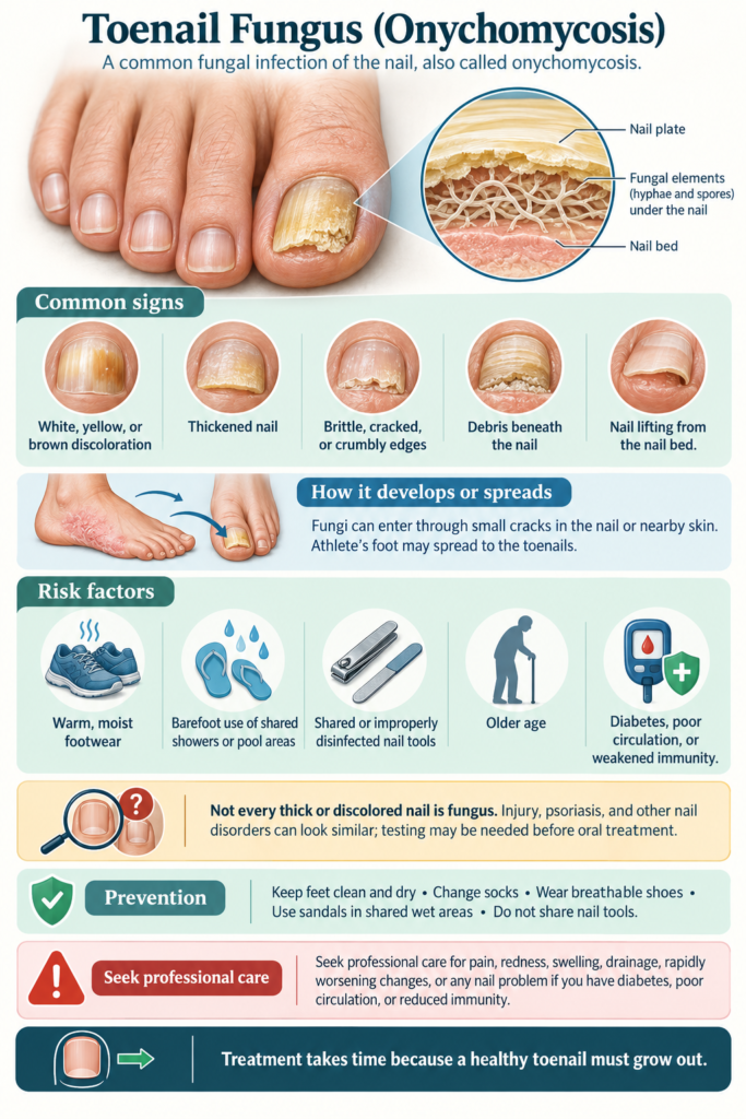
Toenail Fungus
A thickened, discolored nail peeking through your sock can signal an infection that takes far more patience to resolve than it did to acquire. Onychomycosis, the medical term for toenail fungus, is not a superficial cosmetic nuisance but a persistent invasion of the nail bed and plate by dermatophytes, yeasts, or nondermatophyte molds. Your risk climbs quietly over the years as minor trauma to the nail, slower nail growth, and accumulated exposure to damp environments create an open door for pathogens. Public health data consistently point to a global prevalence hovering around 10% of the general population, with that number spiking to roughly 20% for adults past age 60 and to nearly half of all individuals over 70, making age the single strongest predictor.
A warm, dark shoe interior mimics the conditions of a laboratory incubator, precisely what dermatophytes like Trichophyton rubrum need to digest keratin. You often pick up these fungi through direct skin contact with infected flakes shed by another person onto locker room floors, pool decks, or shared towels, though a family history of athlete’s foot can point to genetic susceptibility in how your immune system recognizes the organism. Symptom progression follows a predictable arc, beginning with a white or yellow dot under the tip of the nail that can spread into crumbly debris, a lifted nail plate, and emanating odor if ignored. An estimated 90% of toenail infections trace back to a preceding, chronically untreated case of tinea pedis between the toes, underscoring how skin and nail infections feed each other in a constant cycle of reinfection.
Crowded living conditions, military service, and participation in communal sports amplify transmission rates well above the average, as do health conditions that compromise circulation to your feet, including diabetes and peripheral vascular disease. A 2018 review of epidemiological surveys across North America and Europe confirmed that men are affected roughly 1.5 to 2 times more often than women, a gap often attributed to higher rates of repeated nail microtrauma and occupational footwear use. Treatment landscapes now range from daily terbinafine tablets that achieve a mycological cure in approximately 70% of cases to newer topical agents like efinaconazole that use low surface tension to slip under the nail plate without pills. For stubborn infections and those resistant to first-line therapy, clinicians can pair oral antifungals with periodic debridement, laser-generated heat to damage fungal hyphae, or surgical nail avulsion when a thickened nail refuses to respond to medication alone.
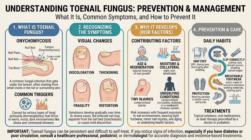
Key Takeaways:
- Dermatophytes, a group of fungi that thrive on keratin, cause the vast majority of toenail infections. The anatomy of the foot creates a warm, moist environment inside shoes that acts as an ideal incubator for these organisms, while microscopic trauma to the nail plate from athletic activity or tight footwear provides a critical entry point.
- Infected nails follow a predictable progression from a small white or yellow spot under the tip to full-blown thickening, crumbling, and debris accumulation under the plate. The nail often separates from the nail bed, a condition called onycholysis, which allows secondary bacterial and yeast infections to move into the newly created space.
- Prevalence rises sharply with age, affecting roughly 10% of the general population but up to 50% of people over 70. Circulation deficits, slower nail growth, and cumulative decades of fungal exposure mean that an active infection in an elderly patient usually requires targeted clinical treatment to resolve.
- Oral terbinafine remains the pharmacological gold standard, achieving a complete cure in approximately 76% of cases over a 12-week course. The drug reaches to the nail matrix through the bloodstream, unlike topical alternatives. It then creates a fungicidal reservoir that stays in the nail for months after the last dose is taken.
- Laser therapy targets the fungus with selective photothermolysis, heating the nail bed to temperatures that denature fungal proteins while theoretically sparing surrounding tissue. Clinical outcomes vary substantially based on the device wavelength and number of sessions, with most protocols requiring three to five treatments spaced four to six weeks apart at an out-of-pocket cost often exceeding $1,000 total.
- Homeopathic approaches, such as preparations of Silicea, Antimonium crudum, or Graphites, are selected based on constitutional symptom profiles, including nail brittleness, discoloration pattern, and surrounding skin texture. These remedies sit outside mainstream clinical evidence, as randomized controlled trials showing a cure rate competitive with terbinafine have not been published.
- Recurrence prevention hinges on footwear hygiene and environmental decontamination, not simply treating the nail in isolation. Spores persist for months in shoes, socks, and shower floors, making a protocol that includes UV shoe sanitizers, frequent sock changes to reduce moisture, and a dedicated pair of shower sandals in gyms and locker rooms a practical imperative for long-term clearance.
Types and Causes of Toenail Fungus
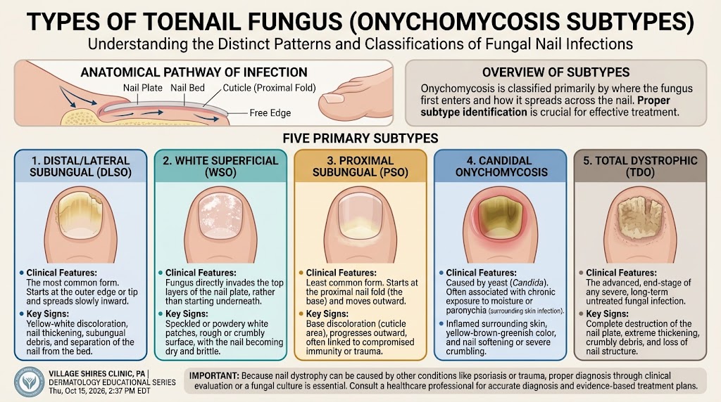
Types of Toenail Fungus
Dermatophytes are responsible for the vast majority of toenail fungal infections, a condition medically termed onychomycosis. Within this group, Trichophyton rubrum stands as the most common pathogen isolated from infected nail samples worldwide, typically initiating an invasion from the hyponychium under the nail’s distal edge. A separate subtype, often more challenging to treat, involves a superficial white onychomycosis where the fungus directly colonizes the nail plate’s surface, leaving a chalky, crumbly texture that you can easily scrape off. This form is frequently linked to Trichophyton mentagrophytes and tends to progress more rapidly in tropical environments.
Nondermatophyte molds cause a smaller but still important number of infections, especially in people who have had repeated trauma to their nails or who already have a weak immune system. Scopulariopsis brevicaulis is a notable example, often appearing after a minor injury and producing a distinctive yellow-brown discoloration along the nail’s edge without the typical thickening of dermatophyte cases. These molds generally fail to produce the keratinolytic enzymes that make dermatophytes so invasive, so they usually exploit a nail already damaged by disease, pressure from footwear, or a prior fungal infection.
Yeast-derived infections, primarily from Candida species, present an entirely different clinical picture that frequently involves the surrounding proximal nail fold before the nail itself. Chronic mucocutaneous candidiasis can manifest with significant paronychia, where the cuticle separates, allowing the yeast to penetrate the nail matrix and distort growth permanently. Your risk for this candidal subtype increases substantially if your occupation requires frequent handwashing, though for toenails, moisture trapped by occlusive footwear creates a similar permissive microenvironment for Candida albicans to proliferate around the toe’s proximal groove.
| Fungal Category | Primary Pathogen | Distinguishing Feature |
|---|---|---|
| Dermatophyte | Trichophyton rubrum | Distal subungual invasion with subungual debris |
| Dermatophyte (Superficial) | Trichophyton mentagrophytes | Chalky white, superficial nail plate colonization |
| Nondermatophyte Mold | Scopulariopsis brevicaulis | Opportunistic infection after nail trauma |
| Yeast | Candida albicans | Proximal nail fold inflammation and matrix damage |
Causes of Toenail Fungus and Risk Factors
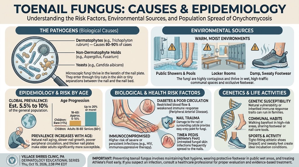
A warm, dark, and moist setting inside your shoe creates the ideal incubation chamber for fungal spores to germinate and penetrate the nail apparatus. Walking barefoot around public pools, locker rooms, and communal showers directly transfers these resilient spores, particularly the ubiquitous Trichophyton species, from contaminated wet floors to the spaces between your toes. Running downhill or wearing narrow toe boxes causes microtrauma to the nail’s attachment point, allowing a separation that spores exploit, while your nail’s slow growth rate hinders the body’s ability to mechanically expel the advancing fungal front.
Circulatory and metabolic conditions dramatically amplify your susceptibility to persistent onychomycosis. Peripheral vascular disease reduces the delivery of immune cells to the distal extremities, leaving the toenails without an effective surveillance mechanism against invading hyphae (the branching filaments of the fungus). Meanwhile, diabetes mellitus creates a dual threat: diminished sensation prevents you from noticing minor cuts or pressure points, while elevated glucose in the nail keratin provides an enriched nutrient source for the fungus. A history of athlete’s foot, or tinea pedis, frequently precedes nail plate infection because the skin infection disrupts the protective barrier at the toe web, allowing dermatophytes to advance into the nail bed.
The structural integrity of your nail itself dictates how easily a pathogen can establish a foothold. Psoriatic nail disease inherently weakens the nail bed’s adhesion, causing onycholysis that leaves a gap where debris and spores collect without resistance. Advancing age further lowers the barrier as your natural nail growth slows, potentially dropping to half its youthful speed by the seventh decade. Repeated athletic impact, such as from tennis or soccer, where the nail repeatedly strikes the shoe’s interior, generates microscopic bleeding under the plate, and a resolving subungual hematoma serves as a protein-rich substrate for opportunistic molds like Aspergillus species to colonize. Family history cannot be ignored as a contributing factor, with certain human leukocyte antigen types showing a genetically weaker defense against dermatophyte invasion.
Signs and Symptoms of Toenail Fungus
Common Signs
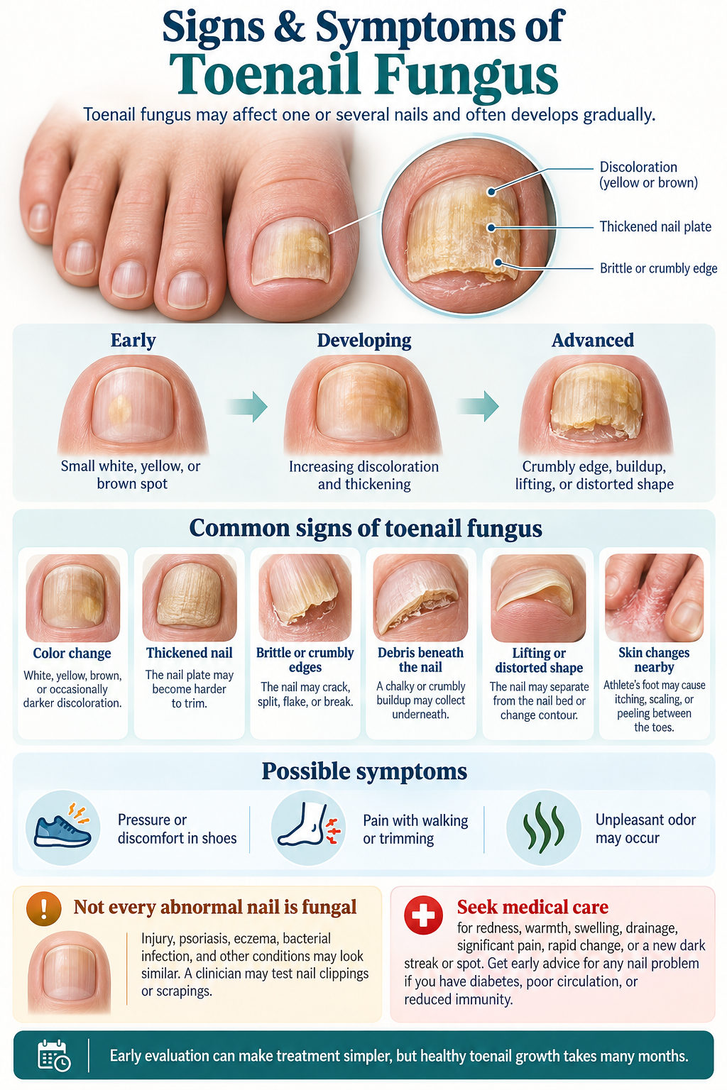
One of the earliest clues you might notice is a shift in your nail’s natural hue, typically beginning at the distal edge and marching toward the cuticle. A chalky white or dull yellow spot appears, often mistaken for a minor bruise, but unlike a bruise it persists and spreads incrementally across the nail plate. Subungual hyperkeratosis is the buildup of crumbly debris under the nail. This slowly lifts the keratin shield from its bed, leaving a visible gap where the dermatophyte colony lives. Thickened nails present a second unmistakable hallmark, as the fungal invasion disrupts the normal layering of keratinocytes and forces the nail to build upon itself in a disorganized, opaque mass. Trimming such a nail becomes a physical challenge with standard clippers, a detail frequently reported to podiatrists during initial consultations.
Distortion of the nail contour proceeds through several recognizable stages, ranging from a slight longitudinal ridging to a pronounced upward prying known as onycholysis. Partial separation from the nail bed leaves a hollowed pocket where moisture and secondary bacteria can accumulate, changing the nail’s architecture permanently if left unaddressed. The surface of an infected nail may develop powdery white islands of superficial fungus, a form that remains confined to the dorsal plate and responds more readily to topical agents than deeper infections do. Some individuals detect a faint, musty odor emanating from the affected toe, a byproduct of keratin degradation that most perceive as socially distressing. When multiple toes on the same foot present with crumbly, discolored edges, the pattern often reflects a long-standing tinea pedis infection that migrated from the plantar skin into the nail apparatus.
Texture changes offer additional diagnostic breadcrumbs during a clinical exam, particularly the brittle fragmentation that causes the nail to shatter like dry chalk under minor compression. These friable edges snag on socks and bedsheets, sometimes prompting a person to modify their footwear choices or avoid barefoot activities altogether. Proximal subungual onychomycosis, though rarer in healthy adults, initiates at the base of the nail with a milky discoloration near the lunula that signals an internally seeded infection in immunocompromised hosts. A darkened nail that mimics a subungual hematoma can also stem from melanonychia induced by certain nondermatophyte molds, including *Scytalidium dimidiatum*, which produce a brownish-black pigment that simulates dried blood under the plate. Nail experts distinguish this fungal melanonychia by its longitudinal band pattern and the absence of any remembered trauma event.
- Debris scraped from the nail often emits a cheesy, keratinous scent.
- On a dermatoscopic view, jagged proximal edges within white streaks suggest ongoing fungal advance.
- Secondary bacterial overgrowth can add a greenish cast from pseudomonas co-infection.
- Pain from a thickened nail pressing against the toe box may alter your gait subtly.
- Fungal reservoirs in neighboring skin scales constantly reseed the nail matrix.
When to Seek Care
Foot pain that extends beyond a cosmetic nuisance requires a prompt podiatric evaluation, particularly when a thickened nail plate excavates into the soft tissue of the lateral nail fold. Ingrown nail fragments driven by fungal deformity can lacerate the sulcus and open a portal for cellulitis, a rapidly spreading bacterial infection identifiable by streak-like redness and local heat. Diabetic neuropathy masks these warning stimuli, so a seemingly minor fungal nail becomes an urgent clinical priority when it abuts a neuropathic ulcer, a combination implicated in the cascade leading to lower extremity amputation. An estimated 15% of diabetic foot complications that result in hospitalization begin with a fungal nail acting as the primary saboteur of skin integrity. Visual changes coupled with any discharge, whether serous or purulent, signal the transition from a chronic fungal state to an acute mixed infection that demands culture and systemic antibiotic coverage alongside antifungal therapy.
The decision to consult a provider should also hinge on the infection’s progression rate and its impact on daily function, two metrics that self-treatment with over-the-counter lacquers rarely halts once the matrix is involved. A nail that completely detaches after minor stubbing, hanging by a narrow proximal hinge, often benefits from professional debridement and sampling for potassium hydroxide microscopy to rule out total dystrophic onychomycosis. Sharp, non-remitting pain while standing or wearing closed shoes indicates that the subungual debris load has reached a point where it splints the nail bed against the terminal phalanx, a mechanical irritation that oral terbinafine or itraconazole can ultimately relieve. Virtually every guideline from the British Association of Dermatologists emphasizes that laboratory confirmation via periodic acid-Schiff staining should precede long-term oral therapy, a step that distinguishes true onychomycosis from psoriatic nail pitting, lichen planus, or traumatic onychodystrophy.
Recurring fungal flares that alternate between remission and relapse despite meticulous topical adherence may indicate an unrecognized dermatophyte reservoir in a household contact or a contaminated grooming tool. Refractory cases in children and adolescents, though less common epidemiologically, warrant a broader immunological workup because persistent dermatophyte invasion can unmask a subtle T-cell deficiency. Certain systemic conditions such as peripheral arterial disease compound the risk of a mixed fungal-bacterial progression, and the altered circulatory status converts an otherwise indolent nail infection into a limb-threatening scenario. A dermatologist can biopsy the nail unit when the pigment distribution raises suspicion for acral lentiginous melanoma, a malignancy that onychomycosis can mask or mimic in its early radial growth phase. Nail plate avulsion performed in-office yields a specimen for histopathology and simultaneously reduces the fungal biomass, accelerating the clinical response to subsequent topical or oral regimens.
Epidemiology and Impact of Toenail Fungus

Patterns of occurrence
Global estimates place the prevalence of toenail fungus, medically termed onychomycosis, at approximately 10% of the general adult population, positioning it as the most frequent nail disorder diagnosed in clinical practice. Rising incidence correlates directly with advancing age, affecting roughly 20% of individuals over 60 and almost 50% of those past 70. General medical clinics report it in about 8% of patients, yet the figure soars to nearly 30% in elderly care settings and can exceed 30% among those with diabetes. Dermatophyte molds belonging to the genus Trichophyton, particularly Trichophyton rubrum and Trichophyton interdigitale, drive the overwhelming majority of cases, responsible for up to 90% of infections worldwide according to mycology findings.
Geographic distribution shifts with climate and localized pathogens, not just host factors. Challenging, chronic infections in tropical nations frequently stem from Scytalidium dimidiatum, a non-dermatophyte mold rarely implicated in temperate regions like the UK, where dermatophyte patterns dominate. Shared household and communal spaces act as powerful transmission hubs; walking barefoot on contaminated locker room floors, pool decks, and hotel bathroom tiles deposits infective keratinous material onto your feet. Nails traumatized by ill-fitting footwear or repetitive athletic stress, such as that seen in long-distance runners, exhibit microscopic separations in the nail plate that allow fungal spores to invade the nail bed directly.
A stark anatomical disparity explains why your toenails suffer 25 times more infections than your fingernails: the slow, keratin-rich growth environment inside shoes creates trapped heat and humidity ideal for dermatophyte proliferation. Genetic predisposition further refines individual risk, as multiple family members within one household frequently share a molecular susceptibility to chronic dermatophyte invasion. Diabetic populations face an amplified threat, with documented prevalence rates between 26% and 34%. Reduced peripheral circulation, compromised immune surveillance, and high glucose concentrations in the nail bed provide a nutritionally rich substrate that accelerates fungal colonization well beyond rates seen in the non-diabetic population.
Effects on nail health and daily life
Left untreated, the infection moves from the distal edge under the nail plate toward the matrix, progressively replacing healthy keratin with crumbly, hyperkeratotic debris that thickens the nail to several times its normal dimension. The resultant disfigurement often traps you in a cycle of embarrassment: a 2022 survey of dermatology patients in the British Journal of Dermatology found that 74% of women and 63% of men reported significant shame over their nail appearance, deliberately keeping their feet hidden in sandals, pools, and intimate settings. Clinical assessment using the Dermatology Life Quality Index validates this psychosocial burden, with onychomycosis patients frequently scoring in the moderate-to-severe impact range, driven by the fear of contamination and unsightly discoloration.
Physical discomfort evolves from a cosmetic concern into a biomechanical safety hazard when neglected. Grossly thickened nails press painfully against the toe box of standard footwear, generating focal pressures that mimic the sensation of a pebble under the toe. Over time, this altered gait places additional load on your hips, knees, and lower spine, contributing to a measurable reduction in postural stability. Cohort studies of elderly individuals with onychomycosis demonstrate a statistically significant increase in fall risk, a danger compounded by the fact that the deformed nail can lacerate adjacent toes or create a sharp edge that snags on bedding, leading to bleeding and bacterial superinfection.
Disease burden magnifies dramatically for the immunocompromised individual, where a breached nail barrier becomes a functional portal of entry for pathogens. Bacterial cellulitis, documented as a direct complication in roughly 10% of diabetic onychomycosis cases, can develop within hours of a minor skin fissure next to an infected nail. Severe, disseminated fungal involvement in patients on chemotherapy or biologic immunosuppressants for rheumatoid arthritis can culminate in complete nail shedding that never regenerates normally. A comparison of distinct populations highlights this unequal distribution of severe outcomes.
| Population | Prevalence | Primary Risk Complication | Functional Impact |
|---|---|---|---|
| Healthy adults under 40 | 2-5% | Recalcitrant cosmetic distortion | Social avoidance and footwear choice limitation |
| Adults over 70 | 45-50% | Thickened nail-induced falls | Gait modification and loss of postural stability |
| Diabetic patients | 26-34% | Bacterial cellulitis entry point | Ulcer formation and potential limb-threatening infection |
| Immunocompromised (transplant, HIV) | Higher than 50% | Proximal subungual invasion and nail shedding | Permanent nail dystrophy and chronic pain |
Medical Treatment Approaches for Toenail Fungus
Prescription and other medical treatments
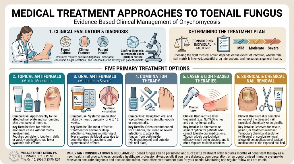
Systemic antifungal drugs form the cornerstone of prescription therapy because they reach the nail bed through the bloodstream rather than relying on topical penetration through the thickened nail plate. Terbinafine, an allylamine, is typically prescribed as a 250 mg daily oral tablet for 12 weeks when treating toenail fungus, and it works by inhibiting squalene epoxidase, an enzyme essential for fungal cell membrane synthesis. A course of itraconazole, a triazole, may instead be dosed continuously at 200 mg daily or as pulse therapy consisting of 200 mg twice daily for one week per month, repeated for three to four months. Laboratory monitoring of liver function is mandated before and during these treatments because terbinafine, in particular, carries a rare but documented risk of hepatotoxicity. Complete cure rates with oral terbinafine reach approximately 55-70% after one year of follow-up, and these figures consistently outperform itraconazole in head-to-head clinical comparisons.
Topical prescription agents provide a non-systemic alternative, though their efficacy depends heavily on the severity of the infection. Ciclopirox 8% nail lacquer, applied daily as a brush-on solution, penetrates the nail plate without systemic absorption and demonstrates a mycological cure rate of roughly 29-36% in mild to moderate distal subungual onychomycosis after 48 weeks. Efinaconazole 10% solution, a newer triazole, achieves higher clearance rates of about 35-45% because its low surface tension aids penetration through the nail to the infection site. Tavaborole 5% solution, a boron-based compound that inhibits fungal protein synthesis, is applied directly to the nail surface and represents another option with a similar safety profile. Your physician may combine an oral antifungal with a topical lacquer to address both the live infection in the nail matrix and the residual fungal elements in the keratin, a dual approach that can reduce reinfection rates, particularly in patients with dermatophytoma or extensive lateral involvement.
Surgical and procedural interventions become relevant when medications alone cannot resolve the condition. Partial or total nail avulsion, performed in a clinical setting under local anesthesia, removes the diseased nail plate to eliminate a large fungal reservoir and permit direct application of topical agents to the nail bed. Chemical avulsion using a 40% urea paste applied under occlusion for 7 to 14 days softens the nail until it can be painlessly debrided, a method often preferred when you have neuropathy or vascular compromise that makes surgical avulsion inadvisable. Debridement itself, whether mechanical by a podiatrist or chemical during at-home care, reduces nail thickness and drug burden but will not cure onychomycosis without concurrent antifungal therapy. These approaches are best suited for infections causing significant pain, those obstructing footwear, or cases where a thickened nail impedes the penetration of topical agents applied over it.
Choosing an effective approach
Building a treatment plan that works for your specific presentation requires matching the infection pattern, your health status, and realistic outcome expectations to the strengths of each option. Dermatophyte infections limited to the distal nail edge in fewer than three nails, for example, often respond adequately to 48 weeks of efinaconazole 10% solution without exposing you to the systemic risks of oral medications. The presence of dermatophytoma, a dense fungal mass within the nail plate visible as a white or yellow streak, signals a barrier that topical solutions rarely overcome alone, making oral terbinafine the more effective primary therapy. Laboratory confirmation through a positive KOH preparation or fungal culture before starting any prescription remains essential because up to 50% of dystrophic nails that appear fungal have alternative causes such as repeated trauma, psoriasis, or lichen planus.
The effectiveness equation also weighs your medication tolerance and co-existing conditions heavily. Oral terbinafine clearance rates in non-dermatophyte molds, particularly Scopulariopsis brevicaulis and Fusarium species, drop well below the 55-70% expected for dermatophytes, sometimes necessitating itraconazole or combination regimens for these less common infections. Your history of congestive heart failure excludes itraconazole use because of its negative inotropic effects, just as active liver disease precludes terbinafine. Duration matters practically as well: it must continue for some months after the nail appears clinically clear because mycological cure often lags behind visible improvement by 6 to 9 months. Reculturing at the end of therapy provides objective proof of eradication rather than relying on the naked eye, a step that distinguishes successful treatment from temporary suppression.
Selecting the single most effective treatment for you specifically hinges on a few concrete factors worth reviewing with your clinician:
- Positive microscopic examination with potassium hydroxide (KOH) preparation confirms a fungal etiology before any prescription fills.
- Terbinafine achieves the highest complete cure rates for confirmed dermatophyte onychomycosis among all oral and topical agents.
- Topical efinaconazole offers a favorable safety profile for patients who cannot take oral antifungals due to drug interactions or hepatic concerns.
- Pulse dosing with itraconazole significantly reduces total drug exposure while maintaining efficacy comparable to continuous therapy.
- Combining a systemic agent with regular professional debridement can shorten the visible recovery time substantially.
- Recurrence rates approach 20-25% within three years, even after successful treatment, making long-term preventive strategies necessary.
Laser, Home Remedies, and Complementary Treatments
Laser and home remedies
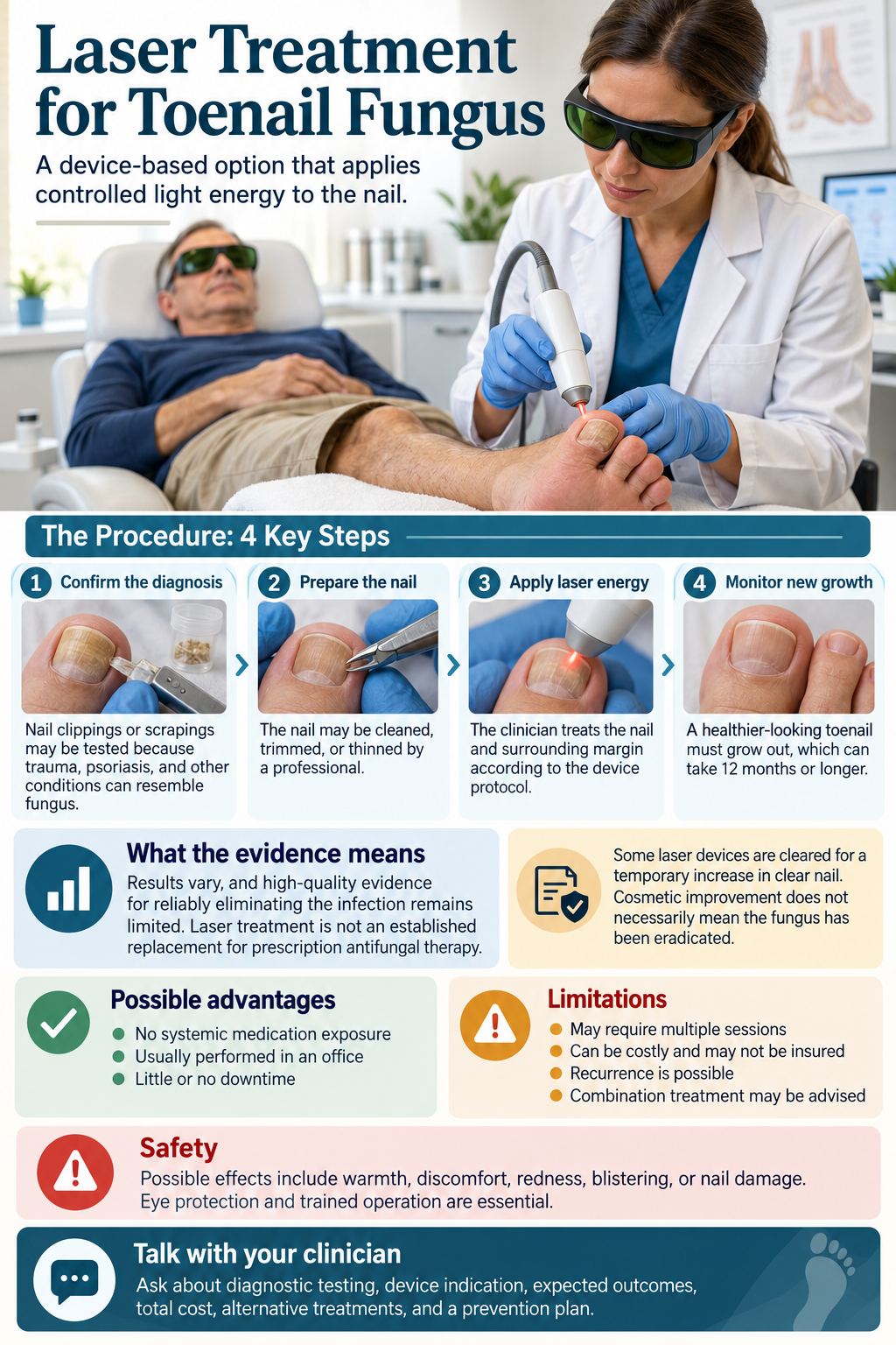
Laser therapy targets the onychomycosis-causing dermatophytes with concentrated light energy that heats and disrupts fungal cells beneath the nail plate without damaging surrounding tissue. A typical treatment course involves two to four sessions spaced several weeks apart, with the Nd:YAG 1064nm and diode lasers being the most commonly employed devices in podiatry clinics. Post-procedure, the nail may appear unchanged for months until healthy regrowth pushes out the infected keratin, and combining laser with a topical antifungal like efinaconazole can modestly improve clearance rates. Insurance rarely covers these treatments, leaving you to manage out-of-pocket costs that range from $300 to $1,200 for a full course depending on your geographic area and the number of nails involved.
Home remedy toenail fungus solutions draw on household items with purported antifungal properties, the most studied being Vicks VapoRub containing camphor, eucalyptus oil, and thymol. Applying a small amount to the affected nail daily for up to 48 weeks led to clinical improvement in a subset of participants in a small 2011 pilot study published in the Journal of the American Board of Family Medicine. Other popular choices include tea tree oil at concentrations of 5 to 10 percent, oregano oil rich in carvacrol, and snakeroot extract, though evidence remains largely anecdotal or derived from in vitro experiments. Snakeroot extract, from the plant Ageratina pichinchensis, showed comparable effectiveness to ciclopirox in one Mexican clinical trial involving 110 patients with mild to moderate disease.
| Approach | Examples | Typical Cost Range | Notable Clinical Data |
|---|---|---|---|
| Laser Treatment | Nd:YAG 1064nm, Diode | $300-$1,200 per full course | Moderate mycological cure rates, boosted by adjunct topical therapy |
| Over-the-Counter Topical | Vicks VapoRub, Tea tree oil 5-10% | $5-$25 per month | 48-week pilot study for VapoRub; Ageratina trial with 110 patients |
Soaking the feet in a vinegar-water solution, typically one part white vinegar to two parts warm water for 15 to 20 minutes daily, aims to create an acidic environment unfavorable to fungal survival. Actual clinical success with this method is poorly documented, and overuse risks skin irritation or maceration that could worsen a concurrent bacterial infection. Baking soda pastes, hydrogen peroxide applications, and garlic extracts circulate in online forums but lack the standardized preparation or dosing needed for reliable physician recommendations. Any soak-based home remedy for toenail fungus must be combined with meticulous drying between the toes, as moisture perpetuates the same environment that encouraged the initial infection.
Natural, holistic, and homeopathic approaches to toenail fungus
Natural holistic treatment for toenail fungus integrates dietary interventions, herbal supplementation, and lifestyle changes that address the internal terrain you provide for fungal proliferation. Candida overgrowth theorists suggest limiting refined sugars, alcohol, and processed carbohydrates while increasing probiotic-rich foods like kefir, kimchi, and unsweetened yogurt to support gut-skin microbial balance. Complementary protocols frequently add oral garlic tablets, caprylic acid derived from coconut oil, or undecylenic acid, a fatty acid with documented anti-Candida properties that has been used in topical antifungal preparations since the 1940s. Crystal-clear data linking these systemic shifts to improved onychomycosis outcomes are absent from robust randomized trials, leaving their use largely in the domain of functional medicine practitioners who monitor progress through repeated nail sampling.
Homeopathic Toenail Fungus Treatment
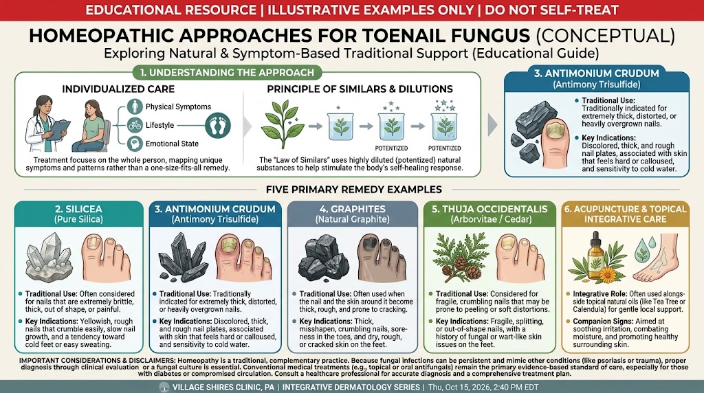
Homeopathic toenail fungus treatment operates on the principle of “like cures like,” employing highly diluted substances that would, in larger doses, produce symptoms mirroring the infection. A classical homeopathic treatment for toenail fungus begins with a constitutional assessment by a licensed homeopath, who considers your emotional patterns, thermal preferences, and coexisting conditions alongside the nail’s actual appearance—brittle, thickened, yellowed, or crumbling. Antimonium crudum sometimes enters the discussion for thick, distorted nails with a tendency to split, while Silicea terra targets individuals with brittle nails and a predisposition to suppuration around the nail bed. Graphites naturalis might be selected when sticky, honey-like discharge and ragged nail edges dominate the clinical picture, particularly in those who tend toward obesity and skin eruptions.
Homeopathic Remedies for Toenail Fungus
Top homeopathic remedies for toenail fungus, prescribed most frequently in Materia Medica texts, include Sepia officinalis for discolored, malformed nails accompanied by a sensation of weight in the pelvic region, and Thuja occidentalis, traditionally associated with warty growths and nail plate ridging. Homeopathic remedies for toenail fungus come as small lactose pellets or liquid drops taken away from meals, with potencies often starting at 6C or 30C and repeated over four to eight weeks before reassessment. Clinicians practicing evidence-based integrative medicine acknowledge that these remedies, prepared through serial dilution beyond Avogadro’s number, act through mechanisms unexplained by current pharmacology and that supportive published evidence consists almost entirely of case series and century-old proving reports. Your decision to pursue homeopathic remedies for toenail fungus should occur within a transparent dialogue where conventional antifungal culture and sensitivity testing proceeds concurrently, preventing avoidable nail dystrophy during periods of non-response.
Customized homeopathic remedies work well for infections, toenail fungus, and nail fungus. A non-invasive and non-toxic therapy option is homeopathy. By addressing the underlying cause, it can totally cure diseased nails. These medications are made from natural materials to treat toenail fungus, nail fungus, and infections.
However, the severity of the infection and the particular remedies selected determine how effective homeopathic treatment is. For the best treatments for your toenail fungus, infection, and nail fungus, you must speak with a licensed homeopath.
The top five homeopathic remedies for toenail fungus
Homeopathy promises to treat nail infections safely, effectively, and without any negative side effects. These are the top five medications for infections, toenail fungus, and nail fungus.
Antimonium crudum, or Ant-c. Antim. crud., is effective in treating nail infections linked to thick, horny skin callosities. Patients with horny warts on their hands and soles are the greatest candidates for this treatment.
Antim. crud. cures itchy, dry skin caused by nail fungus. It is one of the best treatments for infections of extremely brittle and easily broken nails. Their nails grow out of shape and lose their original form. The ideal nails for Antim. crud. are split nails that grow slowly. It can be used when fungus affects the toenail.
Terra Silicea (Sil.)
When it comes to nail fungus, infections, and toenail fungus with pus, silicea is the best treatment. For people with really chilly palms and feet, it works effectively. They have deformed and diseased nails. Silicea is recommended for nail infections accompanied by foul-smelling perspiration from the hands, feet, and armpits. These individuals report experiencing excruciating discomfort in their fingers and toenails. The patient is agitated and restless due to this excruciating discomfort. When it comes to treating inward-grown nails encircled by flesh and blood, silicea is incredibly effective.
Acidum fluoricum (Fl-ac.)
Infection, toenail fungus, and weakened nails can all benefit from fluoric acid. When nails grow quickly, it can be administered. For those with longitudinally ridged nails, this treatment works wonders. There may be painful soreness between the toes in addition to nail changes. It can be administered for nail infections and touch-sore corns.
Rana Bufo (Bufo)
The finest homeopathic remedy for nail fungus, infections, and toenail fungus that causes the nail to turn blue or black is bufo rana. It works well for fungal infections that cause swelling and pus formation in the skin around the nails. Bufo rana is recommended for toenail fungus and nail fungal infections that cause a deep, dull discomfort that travels up to the axilla.
Graphites (Graph.)
When toenails exhibit inward nail growth, graphite guarantees favorable outcomes. Graphites is recommended if your nails have a tendency to ulcerate and the surrounding tissue is inflamed. In cases of deep-seated discomfort that is sensitive to touch, graphites may also be administered. It is administered when the afflicted areas are uncomfortable and sore.
Sometimes there is a nail abnormality that causes the nails to thicken and break readily. Graphite is recommended for nail infections in individuals with fingertip cracks.
Summing up
Selecting an appropriate treatment path for onychomycosis depends heavily on the causative organism, the extent of nail plate involvement, and your personal health profile. Dermatophyte infections, particularly those driven by Trichophyton rubrum, account for the vast majority of cases and often respond to oral terbinafine, which achieves a mycological cure rate exceeding 70% in clinical practice. Non-dermatophyte molds and yeasts like Candida albicans present a different therapeutic challenge, frequently requiring itraconazole or targeted topical formulations after culture confirmation. Your clinician will weigh the risk of drug interactions, especially with statins or certain antidepressants, against the severity of the infection before prescribing a systemic agent.
A growing body of evidence supports the role of physical modalities for patients who cannot tolerate oral antifungals or prefer to avoid them. Laser treatments using Nd:YAG or diode systems generate photothermal effects within the nail bed, achieving temporary clearance in a subset of patients, though recurrence rates remain higher than those seen with systemic therapy. Efinaconazole and tavaborole solutions offer a topical alternative with improved transungual penetration compared to older lacquers, delivering complete cure in roughly 15-18% of mild to moderate distal subungual cases after 48 weeks of daily application. Combining debridement with any topical agent accelerates visible improvement by reducing fungal biomass and permitting deeper drug diffusion into the keratin matrix.
Long-term outcomes hinge on interrupting the cycle of reinfection, which often originates in fomites like footwear, shower floors, and nail grooming tools. A study of household transmission patterns found that sharing unwashed socks or walking barefoot in communal areas doubled the probability of recurrence within two years. Dermatologists now emphasize a preventive strategy that includes treating tinea pedis concurrently, replacing old porous shoes, and using antifungal powders in socks to maintain a dry environment. For immunocompromised individuals and those with diabetes, even a seemingly minor nail infection warrants consistent monitoring, as the breakdown of the nail barrier creates a portal for bacterial entry that can progress to cellulitis.
FAQ

How long does it usually take to get rid of toenail fungus with treatment?
With oral antifungals, a visible healthy nail can replace the old one in 12 to 18 weeks for fingernails, but toenails often take 6 to 12 months because they grow more slowly. The exact time depends on your nail growth rate and how much of the nail was affected at the start of treatment.
What are the most common mistakes people make when treating toenail fungus?
Many people stop treatment too early once the nail looks better, missing that the fungus can survive in the nail bed and cause recurrence. Another common error is failing to treat shoes with antifungal sprays or powders, allowing the infection to be picked up again from contaminated footwear.
Are there any serious risks with toenail fungus medications prescribed by a doctor?
Oral antifungals like terbinafine can rarely cause liver damage, so doctors monitor liver function before and during treatment. Topical prescription solutions have fewer systemic risks but may cause local redness or irritation, and they work best on mild to moderate infections near the tip of the nail.
Do home remedies for toenail fungus really work?
Many home remedies, like vinegar soaks or Vicks VapoRub, show only weak antifungal activity in studies and are unlikely to clear an established infection fully, especially under the nail. They might temporarily improve surface appearance, but medical treatment is needed for a reliable cure in most cases.
How much does laser treatment for toenail fungus cost, and is it worth it?
Laser treatment typically costs a few hundred dollars per session, with multiple sessions often needed, and it is rarely covered by insurance because it is considered cosmetic. While it can temporarily improve nail clarity, success rates vary widely, and it may not clear the infection as reliably as oral medication.
Can toenail fungus go away on its own without treatment?
It is very rare for a toenail fungus infection to resolve on its own, because the fungus is sheltered under the nail, where the body’s immune response is limited. Without intervention, the infection usually worsens over time, spreading deeper into the nail and possibly to other toes.
How can I prevent toenail fungus from coming back after it clears?
Keep feet clean and dry, change socks daily, and wear breathable shoes to reduce moisture, which fungi need to thrive. Also, disinfect or discard old footwear that may harbor spores, and avoid walking barefoot in communal wet areas like locker rooms and pool decks.
Natural Treatment for Toenail Fungus in Philadelphia

Philadelphia Holistic Clinic is the most advanced naturopathic facility in Brotherly Love. Under one roof, different specialists are ready to provide any available natural treatment for toenail fungus.
Our best weapons to fight toenail fungus are
and in each of these areas of medicine, our practitioners are the best.
When you schedule your appointment for a comprehensive holistic evaluation, you will meet homeopathic practitioner Victor Tsan, who will examine you and explain the best treatment approach for your case.
Contact Philadelphia Holistic Clinic at (267) 403-3085 or scan the QR code below to use our online scheduling system.